Mon - Sat : 9:00 AM - 6:00 PM
- +91-8448877746
- sumitneuro@gmail.com
Brain Specialist in India: When to See a Neurologist or Neurosurgeon
A patient guide to choosing the right brain specialist in India: how neurologists and neurosurgeons differ, which symptoms need urgent attention, and which brain conditions may need surgery.
- Vice Chairman, Robotics, Neurosurgery & Spine Services, Max Hospital
- Ex-Professor, JPNA Trauma Center & AIIMS, New Delhi
- MS, DNB, MCh, FACS
- Hon'y President NTSI · President ISPNS
- Keyhole & endoscopic brain and spine surgery
- Honest advice — surgery only when needed
"Brain specialist" is a phrase patients use for any doctor who looks after the brain, but in practice it covers two quite different specialists: the neurologist, who diagnoses and treats brain disorders mainly with medicines and other non-operative therapies, and the neurosurgeon, who is trained to operate on the brain, skull, spine and nerves when surgery is the right answer. Knowing which one to see first, and how urgently, can save precious time, particularly when a symptom is the first sign of a bleed, a tumour or rising pressure inside the skull.
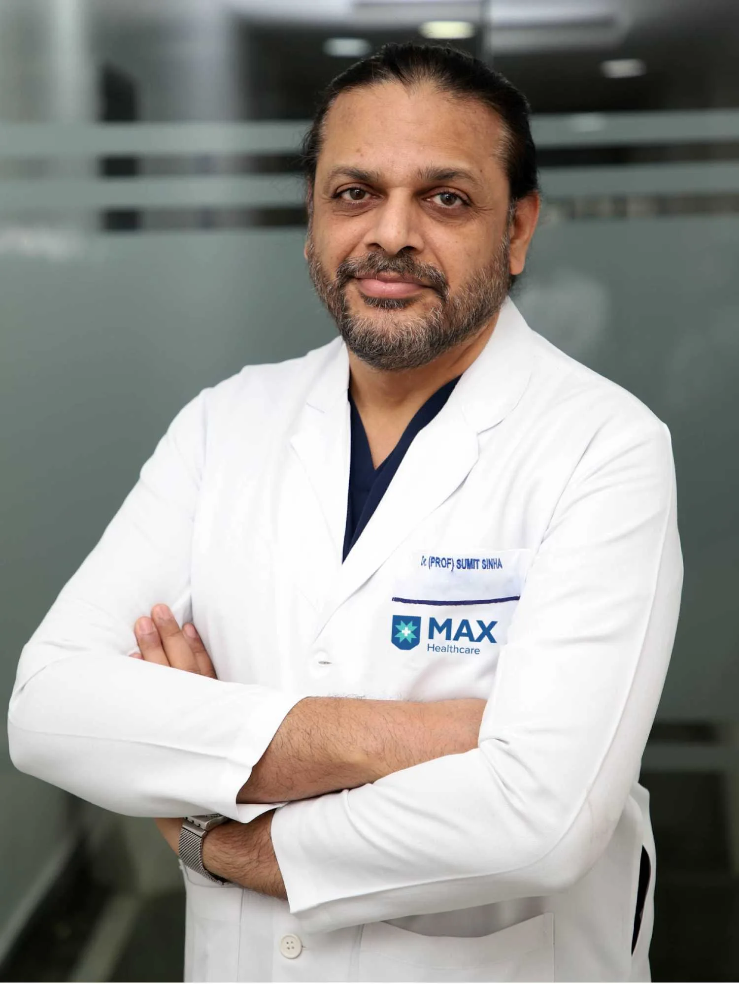
This guide is written by the team of Dr. (Prof.) Sumiet Snha (also known as Dr. Sumit Sinha), an AIIMS-trained neurosurgeon, former Professor of Neurosurgery at AIIMS New Delhi and now Principal Director – Neurosurgery & Spine Surgery at Max Super Speciality Hospital, Dwarka, New Delhi. It explains the warning signs that should never be ignored, the tests a brain specialist is likely to order, and the brain conditions for which surgery may be recommended. It is intended to help you ask the right questions, not to replace a medical examination.
The two specialities share the same organ but different tools. A neurologist completes MBBS, an MD in medicine and then a DM in Neurology; a neurosurgeon completes MBBS, usually an MS in surgery, and then an MCh or DNB in Neurosurgery. Neither is "senior" to the other. In good hospitals they work side by side, and a patient may move between them as a diagnosis becomes clearer. The table below summarises typical reasons to see each, though there is considerable overlap.
| Usually a neurologist first | Usually a neurosurgeon first |
|---|---|
| Migraine and recurrent headaches with a normal scan | A mass, cyst or tumour seen on MRI or CT |
| First seizure or newly diagnosed epilepsy | Seizures that continue despite two or more suitable medicines |
| Stroke prevention and clot-related (ischaemic) stroke | Brain haemorrhage, aneurysm or arteriovenous malformation |
| Parkinson's disease, tremor and movement disorders | Hydrocephalus or a blocked shunt |
| Memory loss and dementia assessment | Head injury with bleeding or skull fracture |
| Multiple sclerosis, neuropathy, muscle disease | Trigeminal neuralgia not controlled by medicines |
If you are unsure, start with whichever specialist you can see soonest. A responsible neurologist will refer you to a neurosurgeon when an operation may help, and a responsible neurosurgeon will refer you back to a neurologist when medical treatment is the better path. Dr. Snha works closely with neurologists for exactly this reason, and many of his brain patients are referred by them.
Some brain symptoms cannot wait for an outpatient appointment. They may signal bleeding, a blocked blood vessel, rapidly rising pressure inside the skull or a serious infection, and outcomes depend heavily on how quickly treatment begins. In these situations the right first step is the nearest hospital emergency department with CT scanning, not a clinic visit or an online search. Once the immediate danger has been assessed, a neurosurgeon can be involved if the scan shows a problem that needs an operation.
Go to emergency immediately if you notice
A sudden, severe "worst-ever" headache that peaks within seconds or a minute; sudden weakness or numbness of the face, arm or leg, especially on one side; sudden difficulty speaking or understanding; sudden loss of vision or double vision; a seizure for the first time or one lasting more than five minutes; confusion or drowsiness after a head injury; or headache with fever, neck stiffness and a rash.
Remember BE-FAST for stroke
- Balance: sudden loss of balance or coordination.
- Eyes: sudden blurred, double or lost vision.
- Face: one side of the face droops when smiling.
- Arm: one arm drifts down when both are raised.
- Speech: slurred or confused speech.
- Time: note the time symptoms began and call for help at once.
The time of onset matters because clot-dissolving and clot-removal treatments for stroke work only within limited time windows. A CT scan will also show whether the stroke is caused by bleeding, which is managed very differently and may need a neurosurgeon.
Many brain conditions announce themselves slowly. A tumour, a chronic subdural haematoma or hydrocephalus may cause symptoms that creep up over weeks, and families often put them down to age, stress or tiredness. These symptoms do not usually need an ambulance, but they do deserve a specialist assessment within days rather than months, particularly when they are new, steadily worsening or combined with one another.
Symptoms to take seriously
- A new headache in someone over 50, or headaches that are worse on waking, with vomiting, or on coughing and straining.
- Gradual weakness, clumsiness or dragging of one leg.
- Personality change, apathy or new difficulty with planning and memory.
- Progressive hearing loss or ringing in one ear, with or without imbalance.
- Gradually narrowing vision or bumping into objects on one side.
- Unexplained changes in menstrual cycle, milk discharge or growth of hands and feet, which can point to a pituitary problem.
- Unsteady walking with urinary urgency and forgetfulness in an older adult.
- Brief episodes of strange smells, déjà vu or staring spells.
None of these symptoms means a serious condition is definitely present; most have harmless explanations. The point is that they warrant an examination and, often, an MRI scan, so that a treatable cause is not missed.
Get an expert opinion before you decide
Share your MRI / CT reports on WhatsApp for a preliminary opinion, or book an in-person / video consultation.
A careful history and neurological examination remain the foundation of diagnosis. The specialist will test strength, sensation, reflexes, coordination, eye movements, speech and memory, and look at the back of the eye for signs of raised pressure. Investigations are then chosen to answer specific questions. More tests are not always better: the aim is to reach an accurate diagnosis with the fewest, most informative investigations.
| Test | What it shows | Typically used for |
|---|---|---|
| CT scan | Bleeding, fractures, large masses, hydrocephalus; very quick | Emergencies, head injury, suspected haemorrhage |
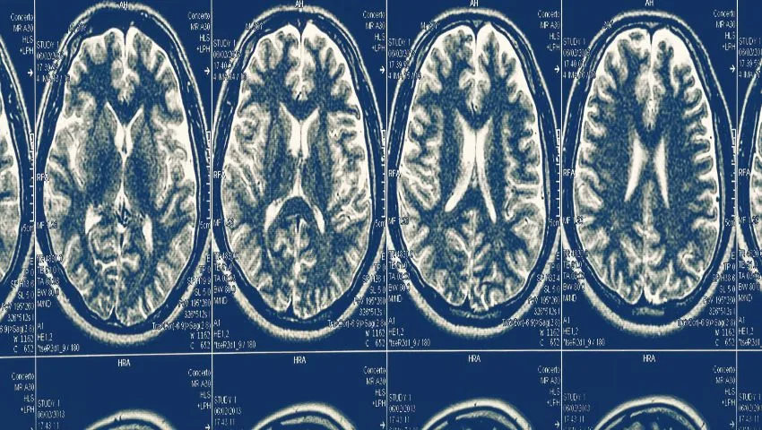
| MRI brain with contrast | Detailed soft-tissue images of the brain and nerves | Tumours, epilepsy, infections, pituitary and hearing-nerve lesions |
| CT or MR angiography | Blood vessels without a catheter | Screening for aneurysms, AVMs and narrowed arteries |
| Digital subtraction angiography (DSA) | The most detailed view of vessels, via a catheter | Planning aneurysm, AVM and bypass treatment |
| EEG and video-EEG | Electrical activity of the brain | Seizures and epilepsy surgery evaluation |
| Advanced MRI (spectroscopy, perfusion, DTI, functional MRI) | Tumour chemistry, blood flow, nerve fibre tracts, speech and motor areas | Characterising tumours and planning safe surgery |
Other supporting investigations
- Hormone blood tests for pituitary tumours.
- Visual field testing when a lesion may be pressing on the optic nerves.
- Audiometry for acoustic neuroma and other tumours near the hearing nerve.
- Neuropsychological assessment before epilepsy or tumour surgery near language and memory areas.
- Lumbar tap test for suspected normal pressure hydrocephalus.
Before travelling for a consultation, it helps to bring the actual scan images on a CD or digital link, not just the printed report. Reports summarise findings, but a neurosurgeon will want to study the images themselves, measure the lesion and look at its relationship to nearby vessels and nerves. Dr. Snha frequently reviews scans shared on WhatsApp before patients decide to travel, so that any additional tests can be arranged efficiently.
Patients are sometimes told that a scan finding is "nothing to worry about" by one doctor and "needs surgery" by another. This is confusing, and it usually reflects the fact that many brain findings sit in a grey zone where both observation and treatment are reasonable. A neurosurgical opinion is especially valuable in the following situations, even when surgery ultimately proves unnecessary:
- Any mass, cyst or abnormal enhancement on a brain MRI.
- An unruptured aneurysm or vascular malformation found by chance.
- Enlarged ventricles (fluid spaces) on a scan, with or without symptoms.
- Epilepsy not controlled after trials of two appropriate medicines.
- Facial pain that is no longer controlled by medicines or causes intolerable side effects.
- Any head injury in a person taking blood thinners, even a minor one.
An early opinion does not commit you to an operation. It gives you an understanding of the natural course of the condition, the options available and the signs that would make treatment more urgent.
Most people who see a brain specialist never need an operation. For some conditions, however, surgery offers the best or only chance of relieving pressure, stopping bleeding, removing a growth or controlling symptoms. The sections below outline the brain conditions Dr. Snha treats most often, the typical symptoms and the surgical options. Each has its own detailed page on this website.
Brain tumours
Brain tumours may start in the brain itself (such as gliomas), in its coverings (meningiomas), in the pituitary gland, or on the nerves at the base of the skull (such as acoustic neuromas). Others spread from cancers elsewhere in the body. Symptoms depend on location: headaches, seizures, weakness, personality change, visual loss or hormonal disturbance. Treatment ranges from observation of small benign tumours to surgical removal, often followed by radiotherapy or chemotherapy for malignant types. Dr. Snha uses microsurgery, neuronavigation and endoscopic techniques, including removal of selected pituitary and skull base tumours through the nose, and has particular experience with acoustic neuroma surgery aimed at preserving facial nerve function.
Brain aneurysm
An aneurysm is a weak, balloon-like bulge in the wall of a brain artery. Many cause no symptoms and are found by chance. If one ruptures, it causes a subarachnoid haemorrhage, typically a sudden, explosive headache with vomiting, neck stiffness or collapse, and this is a life-threatening emergency. Treatment aims to seal the aneurysm from the circulation, either by surgical clipping through a small opening in the skull or by endovascular coiling through a catheter. The choice depends on the aneurysm's shape, size and location and on the patient's condition. Complex aneurysms may need cerebral bypass surgery. Dr. Snha's cerebrovascular work is described on the vascular neurosurgery page.
Arteriovenous malformation (AVM)
An AVM is a tangle of abnormal blood vessels in which arteries connect directly to veins, bypassing the normal capillary network. It is usually present from birth but may first show itself in young adults as a brain haemorrhage, seizures or headaches. Treatment options include microsurgical removal, embolisation (blocking vessels from inside using a catheter) and focused radiation (radiosurgery), used alone or in combination. The right plan depends on the AVM's size, location, drainage pattern and whether it has bled. Some AVMs in critical areas are safest left alone with careful monitoring.
Hydrocephalus
Hydrocephalus is a build-up of cerebrospinal fluid within the brain's fluid spaces, raising pressure inside the skull. In infants it may cause an enlarging head and bulging soft spot; in adults, headaches, vomiting, blurred vision and drowsiness. A slowly developing form in older adults, normal pressure hydrocephalus, causes the triad of unsteady walking, urinary urgency and forgetfulness, and is sometimes mistaken for dementia. Treatment involves diverting the fluid, either by a shunt tube or, in suitable cases, by endoscopic third ventriculostomy, which creates a new internal pathway without implanting a device.
Drug-resistant epilepsy
About two-thirds of people with epilepsy become seizure-free with medicines. For the remainder, whose seizures continue after adequate trials of two appropriate drugs, surgery may be an option if the seizures arise from an identifiable area of the brain. Evaluation typically includes MRI, prolonged video-EEG monitoring and neuropsychological testing. Procedures range from removing a small scar or lesion to anterior temporal lobectomy for mesial temporal sclerosis, and, for some patients, disconnection or neuromodulation procedures. Careful selection is essential, which is why epilepsy surgery decisions are made jointly with neurologists. More detail is available on the epilepsy treatment page.
Trigeminal neuralgia
Trigeminal neuralgia causes brief, electric-shock-like pain in the cheek, jaw, teeth or forehead, often triggered by eating, speaking, brushing the teeth or a light breeze. Many patients first visit a dentist and may even have teeth removed before the diagnosis is made. First-line treatment is medication, usually carbamazepine or a related drug. When medicines stop working or cause unacceptable drowsiness and imbalance, and an MRI shows a blood vessel pressing on the nerve, microvascular decompression can relieve the pressure while preserving facial sensation. Other options include percutaneous procedures and radiosurgery.
Head injury
Head injuries range from mild concussion to life-threatening brain damage. Road traffic accidents and falls are the commonest causes in India. Severity is assessed using the Glasgow Coma Scale and a CT scan. Surgery is needed when a blood clot compresses the brain, when a depressed skull fracture needs elevation, or when brain swelling cannot be controlled by intensive care alone, in which case part of the skull may be temporarily removed (decompressive craniectomy). An extradural haematoma may follow a brief "lucid interval" in which the patient seems well before deteriorating, which is why observation after head injury matters. As President of the Neurotrauma Society of India and a member of the core committee that formulated national guidelines for traumatic brain injury, Dr. Snha has devoted much of his career to this area.
Chronic subdural haematoma
A chronic subdural haematoma is a collection of old blood between the brain and its outer covering. It is most common in older adults and in people taking blood thinners or antiplatelet medicines, and it can follow a fall so minor that the family has forgotten it. Symptoms develop over days to weeks: headache, confusion, drowsiness, slurred speech or weakness on one side, often mistaken for a stroke or dementia. A CT scan confirms the diagnosis. Small collections may be monitored, but symptomatic ones are usually treated by burr-hole drainage, a relatively short operation through small openings in the skull, often followed by rapid improvement. Recurrence is possible, so follow-up scans are part of care.
Other brain and skull conditions treated
Dr. Snha also treats brain abscess, Chiari malformation, hemifacial spasm, moyamoya disease and craniovertebral junction anomalies, as well as congenital brain conditions in children through paediatric neurosurgery.
Conditions & Treatments

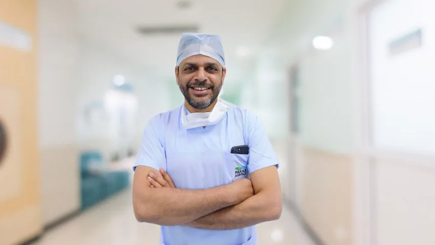
Dr. Snha holds MBBS, MS, DNB and an MCh in Neurosurgery from AIIMS, New Delhi, and is a Fellow of the American College of Surgeons (FACS). After training he served on the AIIMS faculty, becoming Professor of Neurosurgery, and later led neurosurgery at Paras Health, Gurugram, before joining Max Super Speciality Hospital, Dwarka, as Principal Director – Neurosurgery & Spine Surgery. Over more than 25 years he has performed thousands of brain, spine and nerve operations, with a particular emphasis on minimally invasive, endoscopic and keyhole techniques where they are appropriate.
Brain trauma expertise
President of the Neurotrauma Society of India, certified ATLS faculty, and a contributor to national guidelines for traumatic brain injury.
Cerebrovascular interest
Awarded the Young Neurosurgeon Award to attend the 9th International Conference on Cerebrovascular Surgery in Nagoya, Japan.
Teaching and training
Former founder faculty in-charge of the cadaver training and research facility at AIIMS, where surgeons practise complex approaches.
Recognition
Felicitated by Dr. J.P. Nadda, Union Health Minister, and Young Neurosurgeon Award, Times of India Achievers Award 2017.
A first visit usually begins with a detailed history, often with a family member present, since relatives notice changes in memory, behaviour or walking that patients may not. Dr. Snha then performs a neurological examination and reviews your scans on screen, explaining what they show in everyday language. If more information is needed, he will recommend specific tests rather than a broad panel. Where surgery is one of the options, he explains the procedure, its likely benefits, its particular risks in your case and the alternatives, including careful observation. Where surgery is not needed, you will leave with a clear plan and, if appropriate, a referral to a neurologist or other specialist.
For patients outside Delhi
Families from other states and from abroad often start with a remote review. Send your MRI or CT images and reports on WhatsApp to +91-8448877746 or by e-mail to sumitneuro@gmail.com, with a short description of the symptoms. A video consultation can then be arranged to discuss findings and decide whether a visit is required. Max Hospital, Dwarka, is conveniently close to IGI Airport and linked to Gurugram by the Dwarka Expressway, which simplifies travel for outstation and international patients.
When an operation is recommended, the weeks or days beforehand can feel overwhelming. Preparation helps both the patient and the surgical team. Before planned brain surgery, you will usually have blood tests, a heart check (ECG and sometimes an echocardiogram), a chest X-ray and an anaesthesia assessment. Some patients need an updated MRI with a navigation protocol so that the surgeon can plan the safest route to the lesion. Tell the team about every medicine you take, because several common drugs affect bleeding or healing.
Medicines that often need attention before surgery
- Blood thinners such as warfarin, apixaban, rivaroxaban or dabigatran.
- Antiplatelet drugs such as aspirin or clopidogrel.
- Diabetes medicines and insulin, which may need adjusting on the day of surgery.
- Herbal and ayurvedic supplements, some of which can affect clotting.
Never stop a blood thinner on your own. It may have been prescribed for a heart valve, stent or clot, and the timing of stopping and restarting must be agreed between your neurosurgeon and the doctor who prescribed it.
Not sure whether you need surgery?
Many patients improve without an operation. Get a clear, honest plan from Dr. (Prof.) Sumiet Snha.
Recovery depends on the condition treated, the type of operation and the person's health beforehand. Most patients spend the first night in a neuro-intensive care or high-dependency unit, where nurses check consciousness, pupils and limb strength at regular intervals. Many are sitting up and walking within one or two days. Tiredness, mild headache and poor concentration are common for several weeks and gradually settle. Some patients benefit from physiotherapy, speech therapy or occupational therapy, and these are best started early.
Signs to report promptly after discharge
- Increasing headache, vomiting or drowsiness.
- Fever, redness, swelling or fluid leaking from the wound.
- A new seizure, or new weakness, numbness or speech difficulty.
- Clear fluid dripping from the nose after pituitary or skull base surgery.
Driving is usually restricted for a period after brain surgery, and for longer if seizures have occurred; your team will advise based on your situation. Return to office work may be possible within a few weeks after smaller procedures, while heavy physical work and contact sports need a longer interval. Follow-up scans are scheduled according to the diagnosis, and for tumours, the final pathology report guides whether further treatment such as radiotherapy is needed.
Not every brain condition can be prevented, but several important risks can be reduced. Controlling blood pressure lowers the risk of haemorrhagic stroke and aneurysm rupture, and stopping smoking reduces aneurysm growth. Wearing a properly fastened helmet on two-wheelers and a seat belt in cars dramatically reduces the severity of head injuries, which remain a leading cause of death and disability among young Indians. For older adults, preventing falls at home with good lighting, non-slip flooring and grab rails also reduces the risk of subdural haematoma, especially for those taking blood thinners.
Credentials & Leadership Roles
Dr. (Prof.) Sumiet Snha — Minimally Invasive & Endoscopic Neurosurgeon, Spine Surgeon and Peripheral Nerve Surgeon
Awards & Recognition

Felicitated by the Union Health Minister
Swasthya Hindustan Conclave, Zee Hindustan
Honoured by Dr. J.P. Nadda, Union Minister of Health & Family Welfare, for exceptional contribution to neurosurgery and spine surgery. Awards were adjudged by a specialised panel on contributions, success rates and patient outcomes.

Young Neurosurgeon Award
Times of India Achievers Award
Awarded the Young Neurosurgeon Award by a jury of nationally and internationally reputed doctors.

Young Neurosurgeon Award
9th International Conference on Cerebrovascular Surgery, Nagoya, Japan
Selected for the Young Neurosurgeon Award to attend the international conference on cerebrovascular surgery in Nagoya, Japan.
Fellow of the American College of Surgeons (FACS)
American College of Surgeons
Conferred the FACS fellowship - a globally recognised mark of surgical excellence, ethics and quality patient care.
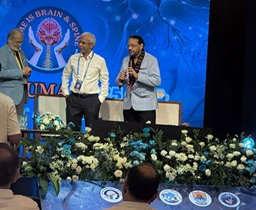
President, Neurotrauma Society of India
33rd Annual Meeting of the Neurotrauma Society of India, Goa
Took over as President of the Neurotrauma Society of India; earlier served as its Secretary.
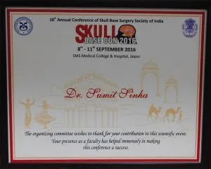
Faculty - Skull Base Surgery Conference
18th Annual Conference of Skull Base Surgery Society of India (Skull Base Con 2016)
Invited faculty for the national skull base surgery conference held at SMS Medical College, Jaipur.
What Patients Say
Congratulations is a small word to wish for Dr. Sumiet Snha for all the hard work you have put into coming so far in life. Your commitment towards become the best neurosurgeon in Delhi & your patients has been always of highest levels. Wish that no height be…
Dr Sumiet Snha for me has been a blessing in-disguise. He explained to me the medical condition in detail along with the treatment options I had. He gave me the right advice and it is because of him today that my slip disc has recovered. He is one of the Top…
My experience with Dr Sumiet Snha has been very pleasant. I have known him for many years and trust him completely. I would highly recommend him for any brain or back problem you may be facing. He always gives the best advice and the right treatment plan for…
Frequently Asked Questions
Not always. "Brain specialist" is an informal term. It may refer to a neurologist, who treats brain disorders mainly with medicines, or a neurosurgeon, who can also perform surgery on the brain, skull and nerves. Many patients need both at different stages.
Most recurring headaches, including migraine, are managed by a neurologist or physician. See a neurosurgeon if a scan shows a structural cause such as a tumour, cyst, bleed or hydrocephalus. A sudden, severe headache needs emergency care before any appointment.
MRI gives the most detailed pictures of brain tissue and is preferred for tumours, epilepsy and many chronic symptoms. CT is faster and better in emergencies, for example after a head injury or suspected bleed. Your specialist will choose based on your symptoms.
No. Some small, slow-growing tumours such as certain meningiomas or acoustic neuromas can be monitored with periodic scans. Others need surgery, radiotherapy, medicines or a combination. The decision depends on tumour type, size, location, symptoms and your overall health.
All brain surgery carries risk, but modern techniques such as microsurgery, endoscopy, neuronavigation and neuromonitoring have made many operations considerably safer than in the past. Your surgeon should explain the specific risks of your procedure compared with the risks of not operating.
It varies widely. After a burr-hole drainage or endoscopic procedure, some patients go home within a few days. After major tumour or vascular surgery, hospital stay may be longer and full recovery can take weeks to months, sometimes with rehabilitation.
Yes. You can send MRI or CT images and reports on WhatsApp to +91-8448877746 or by e-mail to sumitneuro@gmail.com for a preliminary opinion. Video consultations are also available.
Yes. He treats children with hydrocephalus, brain tumours, congenital malformations and head injuries. Read more on the paediatric neurosurgery page.
Book a Consultation With a Brain Specialist
If you or a family member has a worrying brain symptom or an abnormal scan, an early specialist opinion brings clarity. Book an appointment with Dr. (Prof.) Sumiet Snha at Max Super Speciality Hospital, Plot No. 1, Sector-10, Dwarka, New Delhi-110075 (OPD Monday to Saturday), call +91-8448877746, or share your reports on WhatsApp for a preliminary review. Video consultations are available for patients across India and abroad. For any emergency symptom described on this page, please go to the nearest emergency department first. You can also contact the clinic or learn more about Dr. Snha.
- Max Super Speciality Hospital, Plot No. 1, Sector-10, Dwarka, New Delhi - 110075
- +91-8448877746
- sumitneuro@gmail.com