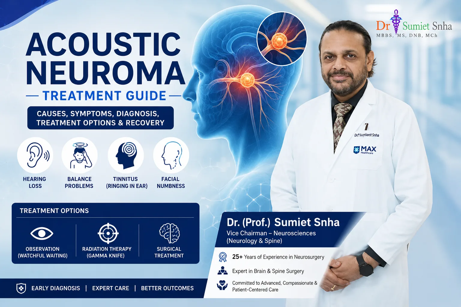
Gradual hearing loss in one ear, a persistent ringing (tinnitus) or a feeling of imbalance are often dismissed as ear infections, wax or simply "getting older". In a small number of people, however, these symptoms are caused by an acoustic neuroma - a benign, slow-growing tumor on the nerve that connects the inner ear to the brain. Although it is not cancerous, an acoustic neuroma can gradually damage hearing and, as it enlarges, press on the facial nerve, the brainstem and the cerebellum. Timely diagnosis and treatment by an experienced team are key to preserving function and quality of life.
Dr. (Prof.) Sumiet Snha, former Professor of Neurosurgery at AIIMS, New Delhi, and Principal Director - Neurosurgery & Spine Surgery at Max Hospital, Dwarka, treats acoustic neuromas of all sizes using microsurgery with continuous nerve monitoring, alongside careful consideration of observation and radiosurgery where appropriate. Many of his patients have shared their recovery stories after acoustic neuroma treatment. This page explains the condition, its symptoms, diagnosis and treatment options in detail.
What Is an Acoustic Neuroma?
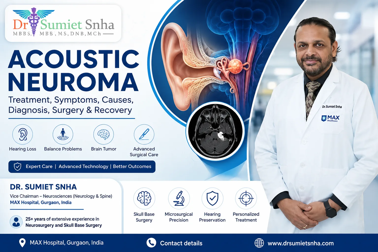
An acoustic neuroma, more accurately called a vestibular schwannoma, is a benign tumor arising from the Schwann cells that form the insulating sheath around the vestibular (balance) nerve. This nerve travels, together with the cochlear (hearing) nerve and the facial nerve, through a narrow bony channel called the internal auditory canal, from the inner ear to the brainstem. The tumor usually begins inside this canal and then grows outwards into the space between the cerebellum and the brainstem, known as the cerebellopontine (CP) angle.
Acoustic neuromas account for a significant proportion of tumors in the CP angle. They usually grow slowly - often a millimetre or two a year - although growth rates vary and some tumors do not grow at all for years. Most occur on one side in adults between 30 and 60 years of age. Tumors on both sides are the hallmark of a genetic condition called neurofibromatosis type 2 (NF2), now often termed NF2-related schwannomatosis.
Anatomy - Why This Location Is Delicate
The CP angle is a small but crowded region. Within a few centimetres lie the nerves for hearing, balance, facial movement and facial sensation, the nerves controlling swallowing and voice, important arteries supplying the brainstem, and the brainstem itself, which controls breathing, heart rate and consciousness. As an acoustic neuroma grows, it stretches and thins the facial nerve - sometimes to a transparent ribbon adherent to the tumor capsule. Separating the tumor from the facial nerve without damaging it is the central challenge of acoustic neuroma surgery, and it is why this operation demands microsurgical expertise and nerve monitoring.
Causes and Risk Factors
For most people with a single acoustic neuroma, no specific cause can be identified. The tumor results from changes in a gene (NF2) that normally suppresses tumor growth in Schwann cells. In sporadic cases, this change occurs only in the tumor cells and is not inherited. In NF2, the change is present in all body cells and can be passed on to children; these patients develop tumors on both sides and often other tumors such as meningiomas and spinal schwannomas. Prior high-dose radiation to the head and neck may slightly increase risk. There is no convincing evidence that normal mobile phone use causes acoustic neuromas.
Symptoms of Acoustic Neuroma
Symptoms develop gradually and depend on the tumor size:
- Hearing loss in one ear - the most common symptom, usually gradual, sometimes noticed first when using the phone on that side. Occasionally hearing is lost suddenly.
- Tinnitus - ringing, buzzing or hissing in the affected ear.
- Imbalance or unsteadiness, particularly in the dark or on uneven ground; true spinning vertigo is less common because the brain compensates for slow nerve loss.
- A feeling of fullness in the ear.
- Facial numbness or tingling - when a larger tumor presses on the trigeminal nerve.
- Facial weakness or twitching - uncommon before treatment, but possible with large tumors.
- Headache, clumsiness and difficulty walking - with large tumors compressing the cerebellum or blocking the flow of cerebrospinal fluid (hydrocephalus).
- Difficulty swallowing, voice change - with very large tumors affecting lower cranial nerves.
Do not ignore one-sided hearing loss
Hearing loss that affects only one ear, or ringing confined to one ear, should always be evaluated by an ENT specialist with a hearing test. If the hearing loss is asymmetric, an MRI is recommended to rule out an acoustic neuroma. Early detection allows treatment while the tumor is small, when the chances of preserving facial function - and sometimes hearing - are highest.
How Acoustic Neuroma Is Diagnosed
- Audiometry (hearing test) - pure-tone audiometry and speech discrimination scores document the degree of hearing loss and help decide whether hearing preservation is realistic.
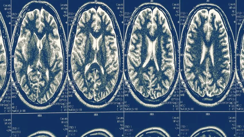
- MRI with gadolinium contrast - the gold standard, detecting even very small tumors within the internal auditory canal and measuring size and relationship to the brainstem.
- Auditory brainstem response (ABR) - may show delayed nerve conduction; used less often now that MRI is widely available.
- Vestibular tests - assess balance nerve function.
- CT scan of the temporal bone - in selected cases for surgical planning.
- Facial nerve assessment - baseline documentation of facial movement.
Tumor Size and Grading
Acoustic neuromas are commonly described by size or grading systems (such as the Koos grading). In simple terms:
Small (intracanalicular)
Confined to the internal auditory canal. Often observed or treated with radiosurgery; hearing preservation may be possible.
Medium
Extending into the CP angle without significant brainstem compression. All three options may be considered.
Large
Touching or compressing the brainstem. Microsurgery is usually preferred.
Giant
Significantly compressing the brainstem and cerebellum, sometimes with hydrocephalus. Requires surgery, often urgently.
Treatment Options for Acoustic Neuroma
There are three main approaches. The best choice depends on tumor size, growth, hearing status, age, general health and the patient's preferences.
1. Observation ("wait and scan")
Small tumors that cause few symptoms, especially in older patients, can be safely monitored with periodic MRI (for example, after 6 months and then yearly). Many small tumors grow very slowly or not at all. Treatment is offered if the tumor grows or symptoms worsen.
2. Stereotactic radiosurgery
Highly focused radiation (for example, Gamma Knife, CyberKnife or linear accelerator-based radiosurgery) delivered in one or a few sessions aims to stop tumor growth rather than remove it. It is suitable for small to medium tumors, typically under about 2.5-3 cm, and for patients who are not fit for surgery. Tumor control rates are high, but radiosurgery does not remove the tumor, and there is a small risk of delayed side effects, including gradual hearing decline and, rarely, facial numbness or weakness. Tumors that continue to grow after radiosurgery can still be operated upon, although surgery may be more difficult.
3. Microsurgical removal
Surgery removes the tumor, providing definitive treatment and immediate relief of pressure on the brainstem. It is the treatment of choice for large tumors, tumors causing brainstem compression or hydrocephalus, cystic tumors, younger patients, and those who prefer removal. The aim is maximal safe removal with preservation of facial nerve function; in selected small tumors with good hearing, hearing preservation may also be attempted.
Surgical Approaches
Retrosigmoid (suboccipital) approach
A craniotomy behind the ear gives excellent access to the CP angle and brainstem. Suitable for tumors of all sizes and allows attempts at hearing preservation.
Translabyrinthine approach
Through the inner ear bone, giving direct access to the internal auditory canal with minimal brain retraction; hearing on that side is sacrificed. Often used in collaboration with an ENT/neurotology surgeon.
Middle fossa approach
From above the ear, for small tumors within the canal when hearing preservation is a priority.
Endoscope-assisted surgery
The endoscope helps visualise tumor remnants in the depth of the internal auditory canal and around corners, improving removal through smaller openings.
Protecting the Facial Nerve - The Heart of Acoustic Neuroma Surgery
The facial nerve controls the muscles of facial expression - closing the eyes, smiling, raising the eyebrows. Losing its function affects appearance, eye protection, speech and eating, and has a major emotional impact. Modern acoustic neuroma surgery places facial nerve preservation at the centre of every decision:
- Continuous facial nerve monitoring (EMG) - electrodes in the facial muscles detect nerve irritation in real time, and a stimulator helps the surgeon locate and map the nerve before dissecting the tumor from it.
- Intracapsular debulking - the tumor is first hollowed out from inside with an ultrasonic aspirator, collapsing it inward so its capsule can be peeled gently away from the nerves.
- High magnification microsurgery - allowing precise identification of the plane between tumor and nerve.
- Judgement over extent of removal - where the tumor is very tightly adherent to the facial nerve or brainstem, leaving a thin layer of capsule (near-total removal) and monitoring it, or treating it with radiosurgery if it grows, can preserve facial function while controlling the tumor.
For small and medium tumors in experienced hands, good facial function is preserved in the majority of patients. With larger tumors the risk is higher, which is one of the strongest reasons to diagnose and treat acoustic neuromas early.
What to Expect During and After Surgery
- Before surgery: MRI review, hearing test, anaesthesia assessment and a detailed discussion of goals and risks.
- Surgery: performed under general anaesthesia with nerve monitoring. Duration depends on the tumor's size and adherence and may range from several hours for smaller tumors to longer for large ones.
- Intensive care: monitoring overnight after surgery.
- Early recovery: dizziness and imbalance are common in the first days because the balance nerve on one side has been removed; the brain gradually compensates.
- Mobilisation: sitting and walking with support usually begin within a day or two; vestibular exercises speed up balance recovery.
- Discharge: typically within a week for uncomplicated cases.
- Follow-up: clinical review and MRI to confirm the extent of removal, and periodic scans afterwards.
Possible Risks and Side Effects
- Hearing loss on the operated side - common, especially with larger tumors or the translabyrinthine approach.
- Facial weakness - temporary or, less commonly, permanent; temporary weakness often recovers over weeks to months.
- Imbalance - usually improves as the brain compensates.
- Tinnitus - may persist.
- Cerebrospinal fluid leak - from the wound or through the nose, occasionally requiring treatment.
- Headache, infection, bleeding and, rarely, injury to other cranial nerves or the brainstem.
- Dry eye - if facial weakness affects eye closure, eye protection with drops, ointment and taping is essential.
Rehabilitation and Recovery
Recovery tips
- Vestibular rehabilitation: specific eye, head and balance exercises help the brain adapt to the loss of balance function on one side.
- Eye care: if blinking is weak, use lubricating drops by day and ointment at night; wear protective glasses outdoors.
- Facial physiotherapy: guided exercises support recovery of facial movement.
- Hearing rehabilitation: options such as CROS hearing aids or bone-anchored devices can route sound from the deaf side to the good ear.
- Activity: gradual return to normal activities over 4-8 weeks; avoid heavy lifting and straining initially.
Most patients return to work and their usual lives within two to three months. Recovery is faster after smaller tumors.
Choosing Between Observation, Radiosurgery and Surgery
For small and medium acoustic neuromas, all three options can be reasonable, and the decision benefits from a frank, individual discussion. Some factors that influence the choice:
- Tumor size and growth: documented growth usually tips the balance toward treatment; large tumors compressing the brainstem generally need surgery.
- Age and health: older patients or those with significant medical problems may favour observation or radiosurgery; younger patients with decades of life ahead may prefer removal.
- Hearing: in small tumors with good hearing, observation or hearing-preservation strategies may be considered.
- Cystic tumors: these can enlarge unpredictably and respond less predictably to radiation, often favouring surgery.
- Patient preference: some people strongly prefer the certainty of removal; others prefer to avoid an operation.
Dr. Snha explains the benefits and trade-offs of each approach clearly, so that the decision reflects both the medical facts and the patient's own priorities.
Acoustic Neuroma and Neurofibromatosis Type 2
Patients with tumors on both sides, or those diagnosed at a young age, may have neurofibromatosis type 2. NF2 requires a specialised, long-term approach because preserving hearing on at least one side is a major priority. Management may include observation, carefully timed surgery, radiosurgery in selected cases, medical therapies used in specialised centres to slow tumor growth, and hearing rehabilitation such as auditory brainstem implants. Genetic counselling is offered to the family.
Why Choose Dr. (Prof.) Sumiet Snha for Acoustic Neuroma?
- AIIMS-trained neurosurgeon - MBBS, MS, DNB, MCh (AIIMS, New Delhi), FACS - with over 25 years of experience in brain and skull base surgery.
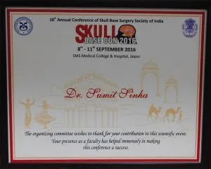
- Former Professor of Neurosurgery at AIIMS, New Delhi, and invited faculty at national skull base surgery conferences.
- Microsurgical removal with continuous facial nerve monitoring and endoscope assistance.
- Balanced, patient-centred advice on observation, radiosurgery and surgery.
- Patient video testimonials include people treated for acoustic neuroma sharing their experience of surgery and recovery.
- Comprehensive care at Max Super Speciality Hospital, Dwarka, with ENT, audiology, physiotherapy and rehabilitation support.
Cost Factors for Acoustic Neuroma Treatment
- Choice of treatment - observation, radiosurgery or microsurgery.
- Tumor size and complexity, which affect operating time.
- Use of neuromonitoring and endoscopic equipment.
- ICU and hospital stay.
- Rehabilitation needs - vestibular therapy, hearing devices.
- Insurance coverage.
Understanding Hearing Loss From Acoustic Neuroma
The hearing loss caused by an acoustic neuroma is called sensorineural hearing loss because it arises from the nerve pathway rather than from the ear canal or middle ear. A characteristic feature is that speech discrimination - the ability to understand words clearly - is often affected more than the simple ability to hear tones. Patients may say that they can hear someone speaking on the phone but cannot make out the words, or that conversations in noisy rooms have become difficult. Because the other ear usually hears normally, many people adapt without realising how much hearing has been lost until they cover the good ear.
The tumor may reduce hearing by pressing directly on the cochlear nerve, by interfering with the blood supply to the inner ear, or by biochemical effects on the cochlea. This explains why hearing may occasionally drop suddenly, mimicking a "sudden sensorineural hearing loss". Any sudden hearing loss in one ear is an urgent reason to see an ENT specialist, and an MRI is usually part of the evaluation.
Balance and Dizziness - How the Brain Adapts
Our sense of balance depends on signals from both inner ears, the eyes and the muscles and joints. An acoustic neuroma slowly damages the balance nerve on one side, and because the change is gradual, the brain usually adjusts - which is why many patients experience mild unsteadiness rather than dramatic spinning vertigo. After surgery, when the remaining balance nerve function on that side is removed, patients typically feel dizzy and unsteady for some days. The brain then recalibrates through a process called central compensation. This is accelerated by moving around, walking and performing specific vestibular exercises; lying still for long periods actually slows recovery. Most people regain good balance within weeks, though some notice mild unsteadiness when tired or in the dark for longer.
Living With Single-Sided Deafness
If hearing is lost on one side, people may find it harder to tell where sounds are coming from and to follow conversations in noisy places. Simple strategies help: sitting with the good ear towards the speaker, choosing quieter restaurant tables, and asking people to face you when speaking. Technology offers further help. CROS hearing aids pick up sound on the deaf side and transmit it wirelessly to the hearing ear. Bone-conduction devices transmit sound through the skull bone to the functioning inner ear. An audiologist can advise on the options best suited to your lifestyle.
Facial Nerve Recovery - What to Expect
If facial weakness occurs after surgery even though the nerve was anatomically preserved, it often improves over weeks to months as the nerve recovers from being stretched and handled. Recovery is usually graded using the House-Brackmann scale, from normal function (grade I) to complete paralysis (grade VI). During recovery, protecting the eye is the most important priority: artificial tears during the day, a lubricating ointment at night, taping the eyelid closed during sleep if needed, and sunglasses outdoors. Facial physiotherapy, including gentle exercises in front of a mirror, can help coordinate the returning movement.
In the uncommon situation where the facial nerve cannot be preserved or does not recover, reconstructive options are available. These include nerve grafting, nerve transfer procedures that connect a nearby healthy nerve (such as the nerve to the masseter or the hypoglossal nerve) to the facial nerve, gold weights to help eyelid closure, and static or dynamic facial reanimation procedures. Dr. Snha's specialised experience in peripheral nerve surgery and nerve transfers is valuable in planning such reconstruction when it is needed.
Headache and Other After-Effects
Some patients experience headaches after surgery, particularly after the retrosigmoid approach. These usually settle over weeks to months and are managed with simple medicines and gradual return to activity. Tiredness is also common for a period after skull base surgery. Tinnitus may persist, improve or occasionally worsen; sound therapy and counselling can help people who find it troublesome. Keeping regular follow-up appointments ensures that any after-effects are recognised and managed early.
Acoustic Neuroma vs Other CP Angle Tumors
Not every tumor in the cerebellopontine angle is an acoustic neuroma. Meningiomas, epidermoid cysts, facial nerve schwannomas and other lesions can occur in the same region and may produce similar symptoms. MRI features help distinguish them - for example, meningiomas often have a broad attachment to the dura, and epidermoids show characteristic diffusion restriction. The distinction matters because treatment strategies and surgical approaches differ. An experienced neurosurgeon reviews the imaging carefully to make the right diagnosis before planning treatment.
Large Tumours and Raised Pressure
Some acoustic neuromas are discovered only when they have become large, because the early hearing loss in one ear went unnoticed or was attributed to other causes. A large tumour can press on the fourth ventricle and block the circulation of cerebrospinal fluid, producing hydrocephalus. Warning signs include worsening headaches, particularly in the morning, vomiting, blurred vision, unsteady walking and increasing drowsiness. In such situations, the pressure inside the head has to be controlled safely before or during tumour removal.
Often, removing the tumour itself restores fluid flow. In other cases, a temporary drain or a procedure such as an endoscopic third ventriculostomy is performed first to relieve pressure, allowing the definitive tumour operation to be undertaken in more stable conditions. With giant tumours, the surgical plan may also prioritise decompressing the brainstem and preserving the facial nerve over complete removal in a single sitting.
Returning to Work, Driving and Travel
Recovery after acoustic neuroma surgery is gradual, and returning to daily roles is best planned in stages rather than all at once. Many people with desk-based jobs resume work part-time once their balance and energy allow, while those in physically demanding work, or work at heights, usually need longer. Tiredness in the first months is normal, and building in rest periods helps.
Practical guidance
- Driving: should wait until balance has recovered, head movements do not bring on dizziness, and any eye or facial symptoms are controlled - your doctor will advise when it is safe.
- Air travel: ask your surgeon before flying, particularly if there has been any fluid leak after surgery.
- Exercise: walking is encouraged early as it speeds balance recovery; swimming and contact sports are resumed only after clearance.
- Eye protection: if blinking is weak, plan for eye drops and glasses while travelling or working at screens.
Preparing for Your Consultation
Deciding between observation, radiosurgery and surgery depends on comparing information collected over time, so bringing complete records makes the first visit much more useful. Please bring all MRI scans on disc along with their reports, especially older scans, which reveal whether the tumour is growing. Audiograms and speech discrimination scores, any balance test results and a list of your medicines are also helpful. Note when your symptoms began and whether they have changed.
If you live outside Delhi or abroad, you can share your reports by WhatsApp on +91-8448877746 or email sumitneuro@gmail.com for a preliminary opinion, and a video consultation can be arranged. Consultations take place at Max Super Speciality Hospital, Dwarka, from Monday to Saturday - book an appointment to plan your visit.
Frequently Asked Questions
Is an acoustic neuroma cancer?
No. An acoustic neuroma is a benign tumor. It does not spread to other parts of the body, but it can cause problems by pressing on nearby nerves and the brainstem as it grows.
Will I lose my hearing after surgery?
Hearing on the affected side is often already reduced, and surgery frequently results in hearing loss on that side, especially for larger tumors. For small tumors with good hearing, hearing-preservation approaches may be possible.
Can acoustic neuroma be treated without surgery?
Small tumors may be observed with regular MRI or treated with stereotactic radiosurgery. Large tumors pressing on the brainstem usually require surgery.
What is the risk of facial paralysis?
With facial nerve monitoring and experienced microsurgery, good facial function is preserved in most patients with small and medium tumors. The risk increases with larger tumors.
How long is the recovery?
Most patients are in hospital for about a week and return to normal activities within two to three months. Balance improves progressively with vestibular exercises.
Can the tumor come back?
After complete removal, recurrence is uncommon. If a small part is left to protect the facial nerve, it is monitored with MRI and treated if it grows.
Why does the tumor cause imbalance?
The tumor arises from the balance nerve. As it damages the nerve, the balance signals from that side reduce, causing unsteadiness until the brain compensates.
Early Diagnosis Protects Your Hearing and Face
If you have hearing loss or ringing in one ear, or have been diagnosed with an acoustic neuroma and are weighing your options, an expert consultation can help you choose the right path. Book an appointment with Dr. (Prof.) Sumiet Snha at Max Hospital, Dwarka, New Delhi. Related pages: brain tumor surgery, microvascular decompression and hydrocephalus.