For a person with a spinal cord injury in the neck (cervical spinal cord injury, or tetraplegia), the loss of hand and arm function is often the most life-changing consequence. Surveys of people living with tetraplegia have repeatedly shown that regaining hand and arm function is their highest priority - above walking - because it determines the ability to eat, write, use a phone, manage personal care, propel a wheelchair and transfer independently. For decades, options to restore upper limb function were limited. Today, nerve transfer surgery offers new hope to many people with cervical spinal cord injury.
Nerve transfers take advantage of nerves that still work - those originating from spinal cord segments above the level of injury - and reroute them to power paralysed muscles controlled by segments below the injury. Dr. (Prof.) Sumiet Snha, Founder Secretary of the Indian Society of Peripheral Nerve Surgery, former Professor of Neurosurgery at AIIMS, New Delhi (including the JPNA Apex Trauma Centre), and President of the Neurotrauma Society of India, brings together deep expertise in spinal cord injury and peripheral nerve surgery to offer nerve transfer procedures at Max Hospital, Dwarka. This page explains how nerve transfers work, who may benefit and what outcomes can be expected.
Understanding Cervical Spinal Cord Injury
The spinal cord in the neck contains segments C1 to C8, each giving rise to nerves supplying specific muscles. The level of injury determines which functions are preserved:
C5 level
Shoulder movement and elbow bending (biceps) are usually preserved; elbow straightening, wrist and hand function are lost.
C6 level
Wrist extension is present, allowing a "tenodesis" grasp, but finger movements and triceps may be weak or absent.
C7 level
Elbow extension is present; finger flexion and fine hand function are limited.
C8-T1 level
Most arm function is present, with weakness of the small hand muscles.
Injuries can be complete (no movement or sensation below the injury) or incomplete (some preserved function). The International Standards for Neurological Classification of Spinal Cord Injury (ISNCSCI) are used to describe the level and completeness precisely.
Why Nerve Transfers Work in Spinal Cord Injury
A key concept is the difference between upper motor neurons (in the brain and spinal cord pathways) and lower motor neurons (the nerve cells in the spinal cord's grey matter at each segment, whose fibres travel in peripheral nerves to muscles). A spinal cord injury damages the upper motor neuron pathways that carry commands down the cord. Below the injury, many lower motor neurons and their peripheral nerves remain intact but are disconnected from the brain's control - these muscles are paralysed but still "wired". Other segments at the level of injury may have lost their lower motor neurons entirely.
In a nerve transfer, the surgeon takes an expendable branch of a nerve that is still under the brain's control (from a segment above the injury) and connects it to the nerve of a paralysed muscle. Because the recipient nerve's pathway to the muscle is intact, the donor nerve fibres can grow into it and reconnect the muscle to voluntary control. This works best when the recipient muscle's lower motor neurons are healthy - which is assessed with electrodiagnostic testing - and when the muscle has not undergone irreversible changes.
Functions That Can Be Restored
Elbow extension
Transfer of a branch of the axillary nerve (e.g. teres minor branch) to the triceps branch of the radial nerve. Elbow extension helps with reaching, pushing a wheelchair, transfers and pressure relief.
Finger and thumb extension (hand opening)
Transfer of the supinator nerve branch to the posterior interosseous nerve, allowing the hand to open to grasp large objects.
Finger flexion (grasp) and thumb flexion (pinch)
Transfer of the brachialis branch (or extensor carpi radialis brevis branch) to the anterior interosseous nerve, restoring the ability to close the fingers and pinch.
Combination procedures
Often, several transfers are performed in one or two stages, on one or both arms, to restore both opening and closing of the hand.
Nerve transfers can be combined with or complemented by traditional tendon transfers, in which working muscles' tendons are rerouted to perform lost functions. Together, these procedures form a comprehensive strategy for upper limb reanimation.
Who Is a Candidate?
- People with cervical spinal cord injury (most commonly C5-C7 levels) who have stable neurological function, typically at least 6 months after injury, when spontaneous recovery has plateaued.
- Availability of suitable donor nerves under voluntary control with good strength.
- Recipient nerves and muscles with intact lower motor neurons, as confirmed by electrodiagnostic studies (muscles respond to electrical stimulation of their nerves).
- Supple joints without significant fixed contractures, or contractures that can be corrected.
- Good general health, skin condition and control of complications such as infections and spasticity.
- Motivation and access to rehabilitation, which is essential for success.
Timing - A Crucial Factor
Timing is one of the most important determinants of success. Surgeons generally wait until neurological recovery has stabilised - often around six months after injury - to avoid sacrificing a nerve that might recover on its own. However, waiting too long is also harmful: in muscles whose lower motor neurons were damaged at the level of injury, the muscle loses its ability to accept new nerve input within about 12 to 18 months. For muscles below the injury with intact lower motor neurons, the window may be longer, but earlier surgery still tends to give better outcomes. The ideal window for many nerve transfers is therefore roughly 6 to 12 months after injury, although selected patients may benefit later. Early referral for assessment - even if surgery is not immediately planned - is strongly encouraged.
Pre-Operative Evaluation
- Detailed neurological examination - muscle-by-muscle strength testing (including hand muscles) and sensory mapping.
- Electrodiagnostic studies (nerve conduction and EMG) - to confirm that recipient nerves and muscles have intact lower motor neurons, and that donor nerves are healthy.
- Assessment of joints and spasticity - range of motion, contractures and tone.
- Functional assessment by occupational therapists - establishing goals and baseline abilities.
- Imaging - MRI of the cervical spine to understand the injury.
- General health review - skin integrity, bladder management, respiratory function and nutrition.
The Surgery
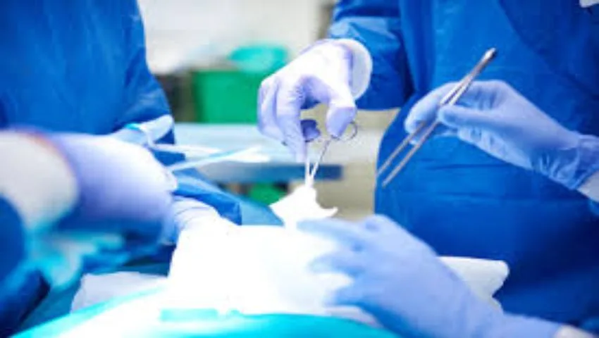
Nerve transfer procedures are performed under general anaesthesia using an operating microscope. Through incisions in the arm or forearm, the donor and recipient nerves are identified and confirmed with a nerve stimulator. The donor branch is divided as close to its muscle as possible, while the recipient nerve is divided as close to its origin as possible, so that the two ends meet without tension. They are joined with extremely fine sutures or fibrin glue. Because the connection is placed near the target muscle, the regenerating nerve fibres have only a short distance to grow. Operations typically take a few hours, depending on the number of transfers, and patients usually stay in hospital for a short period.
Recovery and Rehabilitation
Nerve regeneration is a slow process - roughly one millimetre per day - so recovery unfolds over months:
- First few weeks: the arm is protected, with gentle range-of-motion exercises to keep joints supple.
- 3-6 months: the first flickers of muscle activity typically appear, depending on the transfer.
- 6-18 months: strength progressively improves with targeted exercise.
- Motor re-education: initially, the patient activates the new muscle by trying to perform the donor muscle's original action (for example, trying to bend the elbow to activate finger flexion). With practice, the brain adapts and the new movement becomes more natural.
- Ongoing therapy: occupational therapy focuses on applying new movements to daily tasks - eating, grooming, writing, using devices.
Commitment to rehabilitation is essential. Patients and families should expect a journey of one to two years to reach the full benefit of surgery.
What Results Can Be Expected?
Nerve transfers do not restore normal hand function, but they can provide meaningful, practical improvements. Many patients gain the ability to extend the elbow to reach and push, to open the hand to grasp objects, and to close the fingers and pinch with enough strength for tasks such as holding a cup, feeding themselves, using a phone or keyboard, managing personal care and improving wheelchair skills. These gains can reduce dependence on caregivers and enhance quality of life, confidence and participation in work and society. Results depend on factors such as the level and completeness of injury, time since injury, the health of recipient nerves and muscles, and rehabilitation.
Advantages of Nerve Transfers
- Can restore multiple functions using expendable donor nerves.
- Minimal loss of function from donor sites when properly selected.
- Shorter regeneration distances than traditional nerve grafts.
- Often shorter post-operative immobilisation than tendon transfers.
- Can be combined with tendon transfers for maximal benefit.
- More natural, coordinated movements in many cases.
Risks and Limitations
As with any surgery, there are risks - infection, bleeding, temporary numbness near incisions, and the possibility that the transfer does not produce useful strength. There is also a small risk of weakness in the donor function, which is minimised by selecting expendable donors and testing them carefully. Nerve transfers require time and patience; results cannot be seen immediately. Realistic expectations are discussed in detail before surgery.
Nerve Transfers in Other Conditions
The principles of nerve transfer surgery are widely used in other conditions - notably in brachial plexus injuries, high peripheral nerve injuries such as ulnar and radial nerve palsies, facial nerve paralysis and selected cases of nerve damage after tumors. Dr. Snha's extensive experience with nerve transfers across these conditions informs his approach to spinal cord injury reconstruction.
Comprehensive Care After Spinal Cord Injury
Restoring upper limb function is one part of a broader plan that addresses the many effects of spinal cord injury. This includes management of bladder and bowel function, prevention of pressure sores, respiratory care, control of spasticity and pain, bone health, psychological support, vocational rehabilitation and assistive technology. Early care after injury - including timely decompression and stabilisation of the spine when needed - influences long-term outcomes. Read more on our spinal fracture and spasticity treatment pages.
Why Choose Dr. (Prof.) Sumiet Snha?
- A unique combination of expertise in spinal cord injury and peripheral nerve reconstruction.
- Founder Secretary, Indian Society of Peripheral Nerve Surgery; more than 250 complex nerve and plexus operations.
- President, Neurotrauma Society of India; former Professor of Neurosurgery at AIIMS and the JPNA Apex Trauma Centre.
- Certified AO Spine International and ATLS faculty.
- Expertise in nerve transfers, DREZ and nerve root procedures for spasticity after spinal cord injury.
- Integrated rehabilitation support at Max Super Speciality Hospital, Dwarka.
Cost Considerations
- Number of nerve transfers and whether one or both arms are treated.
- Staged procedures and combination with tendon transfers.
- Electrodiagnostic studies and imaging.
- Hospital stay and long-term rehabilitation.
- Insurance coverage.
Nerve Transfers or Tendon Transfers? Understanding the Choice
People with tetraplegia and their families often ask whether a nerve transfer or a tendon transfer is the better operation. In truth, the two are not competitors but complementary tools, and many reconstruction plans use both. A nerve transfer brings a new nerve supply to a paralysed muscle, so the muscle itself is brought back under voluntary control. A tendon transfer takes a muscle that already works, detaches its tendon and reroutes it to perform a different, more valuable task. Each has its own requirements, timing and recovery pattern, and the right choice depends on which muscles are working, which are paralysed and how long ago the injury occurred.
| Feature | Nerve transfer | Tendon transfer |
|---|---|---|
| What is moved | A working nerve branch to a paralysed muscle's nerve | A working muscle's tendon to a new position |
| Timing | Time-sensitive; best within the first year or so after injury | Can be done years after injury |
| Requirement | Recipient muscle must still respond to electrical stimulation | A strong, expendable muscle must be available to transfer |
| Onset of benefit | Gradual, over many months as nerve fibres regrow | Once healing and retraining are complete, usually within months |
| Immobilisation | Usually brief | Often several weeks of splinting to protect the repair |
| Movements restored | Can reanimate several muscles through one connection | Usually one function per transferred muscle |
Because nerve transfers depend on the health of the recipient muscle, they are generally considered first in people seen within the right window. Tendon transfers remain valuable for those who present later, for muscles whose lower motor neurons have been lost, and to fine-tune function after a nerve transfer has recovered. For example, a patient may have a nerve transfer to restore finger flexion and later a tendon procedure to improve thumb position. Dr. Snha discusses both options openly and, where appropriate, works with hand surgery colleagues so that each arm receives the combination most likely to meet the patient's personal goals.
Measuring Progress After Surgery
Recovery after a nerve transfer is slow, and it is easy to become discouraged when weeks pass with no visible change. Structured measurement helps patients, families and therapists see progress that might otherwise go unnoticed, and it guides decisions about further therapy or additional procedures. At each follow-up visit, the strength of individual muscles is graded using the standard Medical Research Council (MRC) scale, which runs from 0 (no contraction) through a visible flicker, movement with gravity eliminated and movement against gravity, up to 5 (normal power). Reaching grade 3 - movement against gravity - is often the point at which a muscle becomes useful in daily life.
What is assessed at follow-up
- Muscle strength in the reinnervated muscles and in the donor muscles, to confirm donor function remains adequate.
- Electromyography (EMG) in selected cases, which can detect early signs of reinnervation before movement is visible.
- Grip and pinch strength, measured with simple devices once finger movement returns.
- Functional tasks such as picking up objects of different sizes, feeding, writing and using a phone.
- Independence in daily activities, recorded by the occupational therapist using standard questionnaires.
Keeping a simple diary or short monthly video of hand and arm movement at home can also be surprisingly helpful. Small improvements - holding a spoon a little longer, or opening the hand a little wider - are encouraging to see and give the team useful information about how recovery is progressing between clinic visits.
Preparing for Nerve Transfer Surgery: A Practical Checklist
Good preparation makes the surgical admission smoother and helps protect the result. People with spinal cord injury have particular health needs, so planning starts well before the day of surgery. Skin, bladder, chest and positioning all need attention, because a pressure sore or urinary infection around the time of surgery can delay the operation or interfere with early rehabilitation. The team at Max Super Speciality Hospital, Dwarka, reviews these issues at the pre-operative visit and coordinates with the patient's own rehabilitation physician where possible.
- Bring all records - MRI scans, operation notes from the original spinal surgery, previous nerve conduction and EMG reports, and a list of current medicines.
- Check your skin - any pressure area should be treated and healed before surgery; tell the team about past sores.
- Bladder and bowel routine - share your usual catheter or bladder programme so it can be continued in hospital without disruption.
- Breathing - people with higher cervical injuries may benefit from chest physiotherapy and breathing exercises beforehand.
- Blood pressure and autonomic dysreflexia - inform the anaesthetist if you experience episodes of sudden high blood pressure, headache or sweating.
- Equipment - bring your own cushion, splints and any adapted devices you rely on.
- Plan the first weeks at home - since one arm may be temporarily restricted, arrange extra help with transfers and personal care.
It also helps to write down two or three specific goals before the consultation - for example, "to feed myself without a strap" or "to hold a mobile phone". These goals shape which transfers are chosen, which arm is treated first and how therapy is focused afterwards.
The Role of Family and Caregivers
Families and caregivers are an essential part of the reconstruction journey. In the early weeks after surgery, they help protect the operated arm, assist with daily care and make sure the home exercise programme is carried out consistently. Many of the exercises taught by therapists - keeping the shoulder, elbow, wrist and finger joints supple - need a second pair of hands, and stiff joints can limit the benefit of a successful nerve transfer. Caregivers are therefore welcome to attend therapy sessions and learn the correct techniques.
Later, as movement returns, the caregiver's role gradually changes from doing tasks for the person to encouraging them to attempt tasks independently, even when it is slower. This can be emotionally challenging for everyone, but it is how new function becomes part of everyday life. Caring for a family member with tetraplegia over months or years is demanding, and caregivers should also look after their own health, rest and emotional wellbeing. Speaking openly with the treatment team about practical difficulties - transport to therapy, home adaptations or fatigue - allows the plan to be adjusted realistically rather than abandoned.
Incomplete Injuries and Uneven Recovery Between Arms
Not every spinal cord injury follows a neat pattern. In incomplete injuries, some pathways across the injured segment continue to work, and recovery may be patchy - one arm stronger than the other, or some fingers moving while others do not. A common example is central cord syndrome, often seen after a fall in an older person with a narrowed spinal canal, in which the hands are affected more than the legs. In these situations, the priority is usually to allow natural recovery to run its course while keeping joints supple, and to reassess carefully before considering any nerve transfer.
When recovery plateaus, a muscle-by-muscle map of strength shows which functions have returned and which remain absent. Because incomplete injuries may leave several potential donors available, there can be more options - but also a greater need to avoid sacrificing a nerve that is still improving. Surgery may be recommended on one side only, or staged so that the stronger arm is used for daily tasks while the other recovers. These decisions are highly individual and are made jointly with the patient after repeated examination and electrodiagnostic testing.
Travelling for Assessment and Follow-Up
Many people considering nerve transfers live some distance from Delhi, and travel is harder for someone who uses a wheelchair. To reduce unnecessary journeys, a preliminary opinion can often be given by sharing MRI reports, discharge summaries and nerve study results on WhatsApp at +91-8448877746, or through a video consultation. During a video call, the patient can be asked to demonstrate specific movements, which gives a useful first impression of whether nerve transfers are worth exploring. A detailed in-person examination and electrodiagnostic testing are still needed before any surgery is planned.
Max Super Speciality Hospital is in Sector-10, Dwarka, close to IGI Airport and connected to Gurugram by the Dwarka Expressway, which is convenient for patients arriving by air or road. Once surgery is complete, much of the rehabilitation can be carried out with a physiotherapist and occupational therapist near home, following a written programme, with periodic reviews in Dwarka or by video to track progress. To begin, you can book an appointment or write to sumitneuro@gmail.com.
Emotional Wellbeing and Adjusting to Change
Living with a cervical spinal cord injury affects far more than movement. Many people experience grief, frustration, anxiety or low mood in the months and years after injury, and these feelings are a natural response to a life-changing event. Considering reconstructive surgery can bring hope, but it can also stir up worry about whether the operation will work and whether the long rehabilitation will be worthwhile. Acknowledging these emotions openly is an important part of preparing for surgery.
Setting realistic, personal goals helps keep motivation steady during the slow phase of nerve regeneration, when progress may not be visible for several months. Celebrating small milestones - a first flicker of movement, a slightly stronger pinch - makes a real difference. Speaking with a psychologist or counsellor, and connecting with peer support groups of people living with spinal cord injury, can provide practical tips and encouragement that no clinician can fully replace. Family members may benefit from support too.
Ask for help early
If low mood, poor sleep or loss of interest persist for more than a couple of weeks, please mention it at your next visit. Emotional health influences energy, participation in therapy and overall recovery, and support is available as part of comprehensive care.
Frequently Asked Questions
Can nerve transfers restore hand function after spinal cord injury?
In suitable patients with cervical spinal cord injury, nerve transfers can restore useful functions such as elbow extension, hand opening, grasp and pinch, improving independence.
When should nerve transfer surgery be done?
Typically once neurological recovery has plateaued, often around 6 to 12 months after injury. Early assessment is important because delay can reduce the chances of success.
Will I lose any function from the donor nerve?
Donor nerves are carefully selected from expendable branches, so any loss of donor function is usually minimal.
How long before I see results?
The first signs of movement usually appear within 3 to 6 months, with continued improvement over 1 to 2 years with rehabilitation.
Can nerve transfers help people with old injuries?
Some people with older injuries may still benefit, depending on the health of the recipient nerves and muscles. Tendon transfers are another option for later presentations.
Can nerve transfers help me walk?
Nerve transfers for spinal cord injury are primarily used to restore upper limb function. Walking depends on the level and completeness of injury and other rehabilitation strategies.
Is rehabilitation necessary after surgery?
Yes. Dedicated physiotherapy and occupational therapy are essential to retrain the brain and strengthen the reinnervated muscles.
Regain Independence
If you or a loved one lives with a cervical spinal cord injury, an assessment for nerve transfer surgery could open new possibilities. Book a consultation with Dr. (Prof.) Sumiet Snha at Max Hospital, Dwarka, New Delhi. Related pages: brachial plexus surgery, peripheral nerve injuries and spasticity treatment.