A painless lump in the arm, a swelling behind the knee, or a small nodule in the neck that sends an electric jolt down the limb when pressed - these can all be signs of a nerve tumor. Tumors of the peripheral nerves are relatively uncommon, and because they are often small and painless in the early stages, they may be dismissed as a harmless "gaanth" (lump) or misdiagnosed as a cyst or fatty lump. Some are removed by general surgeons without recognising that they arise from a nerve, which can lead to unexpected weakness or numbness afterwards.
The majority of nerve tumors are benign and, in skilled hands, can be removed completely while preserving the nerve's function. A small number are malignant and require specialised, multidisciplinary treatment. Dr. (Prof.) Sumiet Snha, Founder Secretary of the Indian Society of Peripheral Nerve Surgery, former Professor of Neurosurgery at AIIMS, New Delhi, and Principal Director - Neurosurgery & Spine Surgery at Max Hospital, Dwarka, has extensive experience in the microsurgical removal of nerve tumors in the limbs, neck, brachial plexus, pelvis and spine. This page explains the types of nerve tumors, their symptoms, diagnosis and treatment.

What Is a Nerve Tumor?
A nerve tumor is an abnormal growth arising from the cells that make up a peripheral nerve - most often the Schwann cells, which form the insulating sheath around nerve fibres. Tumors can also arise from other components of the nerve or from tissues nearby that press on or invade the nerve. Peripheral nerve tumors can occur anywhere in the body - along the nerves of the arms and legs, the brachial plexus in the neck and shoulder, the lumbosacral plexus in the pelvis, the spinal nerve roots, and the cranial nerves.
Broadly, nerve tumors are divided into:
Benign nerve sheath tumors
Schwannomas and neurofibromas - by far the most common. They grow slowly and do not spread.
Malignant peripheral nerve sheath tumors (MPNST)
Rare, aggressive cancers of the nerve sheath, more common in people with neurofibromatosis type 1.
Other benign nerve lesions
Intraneural ganglion cysts, lipomatous hamartomas, perineuriomas and nerve sheath myxomas.
Non-neural tumors affecting nerves
Lipomas, ganglion cysts, and metastatic or lymphomatous deposits that compress or invade nerves.
Schwannoma
Schwannomas (also called neurilemmomas) are the most common peripheral nerve tumors in adults. They arise from Schwann cells and typically grow as a smooth, rounded, encapsulated mass that pushes the nerve fibres aside rather than growing between them - much like a grape attached to the side of a string. This growth pattern is important because it means that most schwannomas can be separated from the functioning nerve fascicles and removed completely, with a low risk of lasting weakness or numbness when done by an experienced nerve surgeon. Schwannomas are usually solitary. Multiple schwannomas may indicate schwannomatosis or neurofibromatosis type 2. Spinal and intracranial schwannomas - such as acoustic neuromas - belong to the same family; see our acoustic neuroma page.
Neurofibroma
Neurofibromas also arise from nerve sheath cells but, unlike schwannomas, they grow within and between the nerve fibres, intermingling with them. This makes complete removal without affecting nerve function more challenging. Neurofibromas can be:
- Cutaneous - small, soft bumps in the skin, very common in neurofibromatosis type 1 (NF1), usually requiring no treatment unless bothersome.
- Localised intraneural - a discrete swelling within a nerve.
- Plexiform - extensive, rope-like or bag-like growths involving multiple nerve branches, almost always associated with NF1. They can cause disfigurement, pain and functional problems and carry a lifetime risk of transformation into MPNST.
Management of neurofibromas focuses on relieving symptoms and preserving function; complete removal is not always possible or necessary. For plexiform neurofibromas in NF1, targeted medical therapies (MEK inhibitors) have become available in specialised settings and can shrink tumors in many patients.
Malignant Peripheral Nerve Sheath Tumor (MPNST)
MPNSTs are rare soft-tissue sarcomas arising from peripheral nerves. About half occur in people with NF1, often developing from a pre-existing plexiform neurofibroma; others occur sporadically or after radiation. Warning signs that a nerve tumor may be malignant include rapid growth, new or increasing pain (especially at night), a hard, fixed mass and new neurological deficits. Diagnosis requires specialised imaging - often including PET-CT - and a carefully planned biopsy. Treatment involves wide surgical excision with clear margins, frequently combined with radiation and sometimes chemotherapy, coordinated by a sarcoma team. Early recognition is crucial, so people with NF1 should be taught to report changes in their tumors promptly.
Neurofibromatosis - When Nerve Tumors Run in Families
Neurofibromatosis type 1 (NF1)
Café-au-lait skin spots, freckling in the armpits and groin, multiple cutaneous neurofibromas, plexiform neurofibromas, Lisch nodules in the iris, and bone abnormalities. Requires lifelong multidisciplinary follow-up.
Neurofibromatosis type 2 (NF2)
Bilateral vestibular schwannomas, other cranial and spinal schwannomas, and meningiomas. Hearing preservation is a major priority.
Schwannomatosis
Multiple schwannomas, often painful, without the vestibular tumors of NF2.
Genetic counselling
Offered to families, as these conditions can be inherited.
Symptoms of Nerve Tumors
- A lump or swelling along the course of a nerve, often mobile side-to-side but not up-and-down along the nerve's length.
- Electric shock-like tingling radiating along the nerve when the lump is tapped or pressed (a positive Tinel sign) - a characteristic feature.
- Pain - aching or shooting pain along the nerve.
- Numbness or tingling in the area supplied by the nerve.
- Weakness or muscle wasting - usually a late feature of benign tumors; early or progressive weakness may suggest a more aggressive lesion.
- Symptoms mimicking common conditions - a tumor on the sciatic nerve may cause sciatica-like pain; one in the carpal tunnel may mimic carpal tunnel syndrome.
Before removing any lump near a nerve
If a lump produces tingling or electric shocks when pressed, or lies along the path of a major nerve, it should be evaluated with ultrasound or MRI before surgery. Attempting to remove a nerve tumor as if it were a simple cyst or lipoma can damage the nerve and cause permanent weakness or pain. Nerve tumors should be removed by a surgeon experienced in peripheral nerve microsurgery.
How Nerve Tumors Are Diagnosed
- Clinical examination - assessing the lump, its mobility, the Tinel sign, and a detailed neurological examination of the nerve's motor and sensory function.
- High-resolution ultrasound - shows continuity of the tumor with the nerve (the "tail sign") and its internal structure; quick and excellent for initial evaluation.
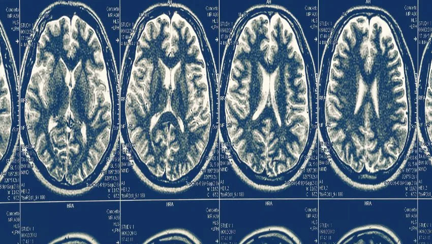
- MRI with contrast - the key test, showing the tumor's size, relation to the nerve, characteristic features such as the "target sign", and features that may suggest malignancy.
- Nerve conduction studies and EMG - to document baseline nerve function.
- PET-CT - when malignancy is suspected, particularly in NF1.
- Biopsy - generally avoided for typical schwannomas (as needle biopsy can damage fascicles and cause pain), but essential when malignancy is suspected; it should be planned by the treating surgical team.
When Do Nerve Tumors Need Treatment?
Not every nerve tumor needs surgery. Small, asymptomatic, typical-looking schwannomas or neurofibromas may be safely observed with periodic imaging. Surgery is usually recommended when:
- The tumor causes pain, numbness or weakness.
- It is growing on follow-up imaging.
- There is diagnostic uncertainty or features suggesting malignancy.
- It causes cosmetic concerns or interferes with daily activities.
Surgical Removal of Nerve Tumors
The goal of surgery for benign nerve tumors is complete removal of the tumor with preservation of nerve function. This requires microsurgical technique and intra-operative nerve monitoring. A typical operation involves:
- Planning: MRI and ultrasound are reviewed to map the tumor and the nerve's course.
- Anaesthesia: general or regional anaesthesia; muscle relaxants are avoided when nerve stimulation is used.
- Exposure: the nerve is exposed above and below the tumor, giving control of the normal nerve on both sides.
- Nerve stimulation and mapping: a nerve stimulator and neuromonitoring identify functioning fascicles on the tumor surface.
- Opening the capsule: in a schwannoma, the outer layer is opened in an area free of functioning fascicles.
- Enucleation: the tumor is gently shelled out, typically attached to a single small, non-functioning entering and exiting fascicle, which is divided.
- Closure: the nerve sheath and wound are closed; no nerve graft is usually needed for schwannomas.
For intraneural neurofibromas, the surgeon removes as much tumor as can be separated safely from functioning fascicles. In the rare situations where a functioning nerve segment must be removed (for example, in malignant tumors), reconstruction with nerve grafts or transfers can restore function.
Results of Nerve Tumor Surgery
For schwannomas, complete removal cures the condition in the vast majority of cases, and most patients have no new lasting deficit; many experience relief of pain and tingling. A temporary increase in numbness or tingling in the nerve's territory can occur in the first weeks and usually settles. Neurofibroma surgery carries a somewhat higher risk to nerve function because of the intermingled growth pattern, which is why the decision to operate on these tumors is carefully individualised. Recurrence after complete removal of a benign schwannoma is uncommon.
Other Lesions Affecting Nerves
Intraneural ganglion cysts
These are fluid-filled cysts within a nerve, most commonly the peroneal nerve near the knee, arising from a nearby joint through a small articular branch. They can cause foot drop. Successful treatment requires not only draining the cyst but also disconnecting the articular branch to prevent recurrence.
Lipomatous hamartoma
A rare condition where fatty tissue grows within a nerve, most often the median nerve, sometimes with enlargement of the fingers. Treatment usually focuses on decompression.
Traumatic neuroma
Not a true tumor, but a painful tangle of nerve fibres after injury - discussed on our nerve injuries page.
Tumors compressing nerves
Lipomas, ganglia, bone tumors and cancerous growths can press on nerves. Removal of the compressing lesion usually relieves symptoms.
Recovery After Nerve Tumor Surgery
What to expect
- Hospital stay: often a day or two for limb tumors; longer for tumors in the plexus or pelvis.
- Wound care: keep the wound clean and dry; stitches are removed in about 10-14 days.
- Activity: gentle movement early; avoid heavy use of the limb for a few weeks.
- Sensory changes: temporary tingling or numbness may occur and usually improves.
- Follow-up: clinical review and, in some cases, imaging to confirm removal.
Nerve Tumors in Challenging Locations
Some nerve tumors arise in anatomically complex areas where experience is particularly important:
- Brachial plexus tumors in the neck and shoulder region lie near major blood vessels, the lungs and the spinal cord. Careful exposure of the plexus and neuromonitoring allow safe removal while preserving arm function.
- Pelvic and lumbosacral plexus tumors may present with leg pain or be discovered incidentally on abdominal scans. They may require abdominal, retroperitoneal or combined approaches.
- Dumbbell tumors extend from inside the spinal canal through the nerve root opening into the neck, chest or abdomen and may need a combined spinal and extra-spinal operation - see our spinal tumor page.
- Sciatic nerve tumors in the buttock or thigh can mimic sciatica from a slip disc and are sometimes missed until a nerve MRI is done.
Dr. Snha's combined expertise in spinal surgery and peripheral nerve surgery is especially valuable for tumors that bridge these regions.
Why Choose Dr. (Prof.) Sumiet Snha for Nerve Tumors?
- Founder Secretary, Indian Society of Peripheral Nerve Surgery.
- Extensive experience with more than 250 complex peripheral nerve and plexus operations.
- AIIMS-trained - MBBS, MS, DNB, MCh (AIIMS, New Delhi), FACS - and former Professor of Neurosurgery at AIIMS.
- Microsurgical removal with intra-operative nerve stimulation and neuromonitoring.
- Expertise across limbs, plexus, pelvis and spine, including dumbbell tumors.
- Multidisciplinary care for neurofibromatosis and malignant tumors at Max Super Speciality Hospital, Dwarka.
Cost Factors for Nerve Tumor Surgery
- Location and size of the tumor.
- Type of tumor and whether reconstruction is needed.
- Imaging and neuromonitoring requirements.
- Hospital stay.
- Further treatment for malignant tumors.
- Insurance coverage.
Understanding Your MRI Report for a Nerve Tumor
MRI reports for peripheral nerve tumors often mention specific signs. The "split fat sign" refers to a rim of fat around the tumor, indicating that it arises within the fat surrounding a neurovascular bundle. The "target sign" - a darker centre with a brighter rim on certain sequences - is typical of benign nerve sheath tumors. The "fascicular sign" describes small ring-like structures within the tumor representing nerve bundles. The "entering and exiting nerve" or "tail sign" shows the nerve continuing into and out of the tumor. Features that raise concern for malignancy include large size, irregular or infiltrative margins, surrounding swelling, areas of necrosis (dead tissue) and rapid change between scans. These features guide whether a tumor can be observed, removed directly, or needs further evaluation for malignancy.
Pain After Nerve Tumor Removal
Many patients experience relief from pain and tingling soon after a schwannoma is removed. Some may have a temporary increase in sensitivity or pins and needles in the nerve's territory for several weeks as the nerve settles after being handled. This usually improves steadily with time, simple pain relief and desensitisation exercises. Persistent or new severe pain is uncommon and should be reported, as it can be evaluated and treated. In patients with schwannomatosis, where pain can be widespread and caused by multiple small tumors, a combination of targeted surgery and pain management strategies is often required.
Living With Neurofibromatosis
People with NF1 or NF2 benefit from coordinated, lifelong care. Regular check-ups monitor known tumors, detect new ones and assess for complications such as learning difficulties, high blood pressure, scoliosis, vision or hearing changes. Patients and families learn the warning signs that warrant urgent review - such as rapid growth, new pain or new weakness in a tumor. Surgery is reserved for tumors causing symptoms or showing concerning changes. Support groups and genetic counselling help families understand the condition and plan for the future. With appropriate follow-up, most people with neurofibromatosis live full, productive lives.
Nerve Tumors of the Hand, Wrist and Forearm
The upper limb is one of the most common sites for peripheral nerve tumors. In the forearm and wrist, schwannomas of the median and ulnar nerves may present as a small, firm, mobile lump that causes tingling in the fingers when pressed. A tumor of the median nerve at the wrist can mimic carpal tunnel syndrome, and if it is not recognised, a routine carpal tunnel release may fail to relieve symptoms. In the hand, small nerve tumors on the digital nerves can cause tender lumps on the fingers. Because the nerves of the hand control fine movement and sensation that are essential for daily activities and work, surgery here demands particular precision. With careful microsurgical enucleation, most patients retain full hand function and return to work quickly.
Nerve Tumors of the Lower Limb
In the leg, tumors most often affect the sciatic nerve and its branches - the tibial and common peroneal nerves - particularly in the thigh and behind the knee (the popliteal fossa). Patients may notice a lump behind the knee, pain radiating into the calf and foot, or numbness on the sole or top of the foot. A tumor of the sciatic nerve in the buttock or thigh can cause pain that closely resembles sciatica from a slipped disc. When spine MRI does not explain a patient's sciatica, a dedicated MRI of the pelvis and thigh or nerve ultrasound can reveal a sciatic nerve tumor. Removing such tumors typically relieves the pain and preserves walking ability.
Head and Neck Nerve Tumors
Schwannomas can also arise from the nerves of the neck - including the vagus nerve, the sympathetic chain, the cervical nerve roots and the brachial plexus. They may present as a painless lump in the side of the neck and are sometimes initially thought to be an enlarged lymph node. Careful imaging distinguishes them from lymph nodes and other neck masses. Because nerves such as the vagus control the voice and swallowing, surgical planning weighs the benefits of removal against the risk to function, and in selected cases, observation may be the best approach for small, asymptomatic tumors.
Monitoring Tumors That Are Not Removed
When a small, benign-appearing nerve tumor is observed rather than removed, a follow-up plan is agreed with the patient. This usually includes a repeat MRI or ultrasound after six to twelve months and then at longer intervals if the tumor remains stable. Patients are asked to report new pain, rapid growth, weakness or numbness between scans. If any of these develop, or if the tumor grows, surgery can be planned at that stage. This approach avoids unnecessary operations for tumors that may never cause problems.
Preparing for Nerve Tumor Surgery
Most nerve tumor operations are planned procedures, which leaves time to prepare properly. A little organisation beforehand helps the surgical team plan the safest approach and makes the hospital stay shorter and more comfortable. At the pre-operative visit, Dr. Snha reviews the imaging in detail, records the strength and sensation in the affected limb as a baseline, and explains the likely incision, the expected hospital stay and the recovery plan. This is also the right moment to raise any concerns about scarring, numbness or time away from work.
- Collect your imaging - bring the actual MRI or ultrasound images on a disc or digital link, not only the written reports, as the images guide the surgical plan.
- Share any previous biopsy or surgery details - if the lump was previously biopsied or partly removed elsewhere, the operation notes and pathology slides or reports are very important.
- List your medicines - especially blood thinners, antiplatelet tablets and diabetes medicines, which may need to be adjusted before surgery on medical advice.
- Arrange help at home - after surgery on the hand or arm, everyday tasks may be awkward for a week or two.
- Plan your return journey - you should not drive yourself home after anaesthesia.
- Check insurance paperwork - pre-authorisation for a planned procedure is easier to complete in advance.
Depending on the tumor's location and size, surgery may be performed under general anaesthesia or, for some small limb tumors, under regional anaesthesia. The anaesthetist discusses the most suitable option, keeping in mind that nerve stimulation during surgery requires that the nerve's responses are not blocked.
Opinion for Patients From Outside Delhi
People with a nerve lump often find themselves unsure whom to consult, and many travel from other cities or countries for specialist advice. To make the first step easier, a preliminary opinion can often be offered by sending MRI reports, ultrasound reports and a brief description of symptoms on WhatsApp at +91-8448877746 or by email to sumitneuro@gmail.com. A video consultation can then be arranged to discuss the findings, the options of observation or surgery, and what further tests might be needed.
If surgery is advised, it is carried out at Max Super Speciality Hospital, Plot No. 1, Sector-10, Dwarka, New Delhi. The hospital's location close to IGI Airport and the Dwarka Expressway makes it accessible for visitors from other states and abroad, and many limb nerve tumor operations require only a short hospital stay. Wound checks and early follow-up can often be coordinated with a local doctor, with later reviews by video. To plan your visit, please book an appointment or contact the clinic.
Wound and Scar Care After Nerve Tumor Surgery
Because many nerve tumors lie in the arms, hands, legs or neck, patients naturally want to know how the scar will look and how to care for the wound. Incisions are usually planned along natural skin creases or in less visible positions where the anatomy allows, and they are kept as short as is consistent with safe exposure of the nerve above and below the tumor. The nerve must be seen clearly on both sides of the lump, so the incision is sometimes longer than the lump itself - a trade-off that protects nerve function.
- First days - keep the dressing clean and dry, and elevate the limb to reduce swelling.
- Stitches - usually removed or checked at around ten to fourteen days, unless absorbable stitches are used.
- After healing - gentle scar massage with a plain moisturiser can soften the scar and reduce tenderness.
- Sun protection - protecting a fresh scar from strong sunlight for several months helps it fade.
- Watch for infection - increasing redness, warmth, discharge or fever should be reported promptly.
Scars typically look pink and slightly raised at first, then flatten and fade over six to twelve months. Some people, particularly those prone to thick scars, may benefit from silicone gel or sheets, which can be discussed at the follow-up visit. A small area of altered sensation around the incision is common and usually becomes less noticeable over time.
Frequently Asked Questions
Are nerve tumors cancerous?
Most nerve tumors, such as schwannomas and neurofibromas, are benign. Malignant nerve sheath tumors are rare, but rapid growth, increasing pain or weakness should be evaluated promptly.
Can a nerve tumor be removed without damaging the nerve?
Yes. Schwannomas in particular can usually be removed completely while preserving the nerve's function, using microsurgery and nerve monitoring.
Why does pressing on the lump cause electric shocks?
Pressure on a tumor arising from a nerve irritates the nerve fibres, producing tingling that radiates along the nerve - a helpful clue to the diagnosis.
Should every nerve tumor be removed?
No. Small, painless tumors that are not growing may be observed. Symptomatic, growing or suspicious tumors are usually removed.
Is a biopsy needed before surgery?
Usually not for typical schwannomas, as MRI features are characteristic. A biopsy is important when malignancy is suspected and should be planned by the treating team.
Can nerve tumors come back?
Recurrence after complete removal of a benign schwannoma is uncommon. Neurofibromas and tumors in neurofibromatosis may recur or develop elsewhere.
What is the recovery time?
Most patients return to normal activities within a few weeks after limb nerve tumor surgery.
Get the Right Diagnosis Before Surgery
If you have a lump that tingles when pressed, or have been told you have a nerve tumor, consult a peripheral nerve specialist before any surgery. Book an appointment with Dr. (Prof.) Sumiet Snha at Max Hospital, Dwarka, New Delhi. Related pages: nerve sheath tumors, peripheral nerve injuries and brachial plexus surgery.