Imagine an electric shock shooting through your cheek every time you brush your teeth, chew, talk, feel a breeze on your face or even smile. For people with trigeminal neuralgia, this is daily life. Often called one of the most painful conditions known to medicine, trigeminal neuralgia can make simple activities like eating and speaking unbearable, leading to weight loss, isolation and depression. Similarly, people with hemifacial spasm experience uncontrollable twitching of one side of the face that may close the eye involuntarily, affecting vision, confidence and social life.
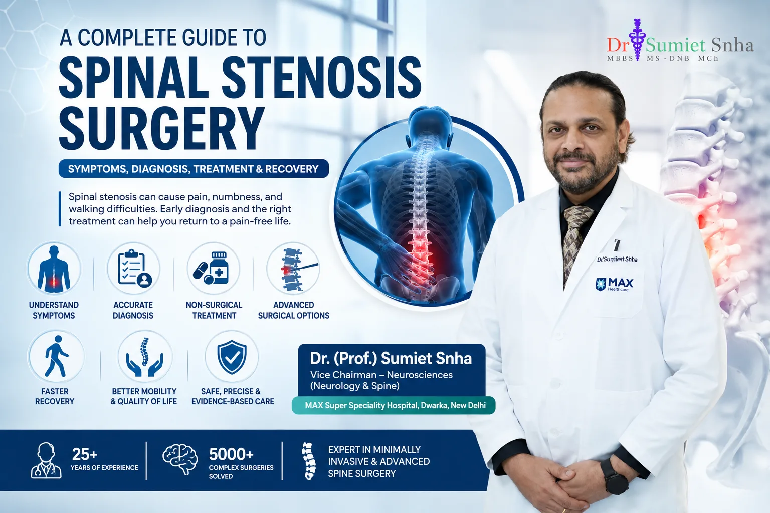
In many patients, both conditions are caused by the same problem: a blood vessel pressing against a cranial nerve where it leaves the brainstem. Microvascular decompression (MVD) is an operation that gently moves the offending vessel away from the nerve and places a small cushion between them. Unlike many other treatments, MVD addresses the cause of the problem while preserving normal nerve function, and it offers the best chance of long-lasting relief in suitable patients. Dr. (Prof.) Sumiet Snha, former Professor of Neurosurgery at AIIMS, New Delhi, and Principal Director - Neurosurgery & Spine Surgery at Max Hospital, Dwarka, performs MVD for trigeminal neuralgia, hemifacial spasm and glossopharyngeal neuralgia using microsurgical and endoscope-assisted techniques.
Understanding the Cranial Nerves and Neurovascular Compression
Twelve pairs of cranial nerves emerge directly from the brain and brainstem. The trigeminal nerve (fifth cranial nerve) carries sensation from the face - the forehead, cheek and jaw - and controls the chewing muscles. The facial nerve (seventh cranial nerve) controls the muscles of facial expression. The glossopharyngeal nerve (ninth cranial nerve) carries sensation from the throat and back of the tongue.
Each of these nerves has a short, vulnerable segment where it leaves the brainstem, called the root entry zone (or root exit zone). Here, the nerve's insulation changes from central to peripheral myelin, and it is particularly sensitive to pressure. With age, arteries in the head tend to elongate and loop, and a small artery - most commonly the superior cerebellar artery for the trigeminal nerve, or the anterior inferior cerebellar artery or vertebral artery for the facial nerve - may come to rest against this segment. The constant pulsation gradually wears away the nerve's insulation, causing nerve fibres to "short-circuit" and fire abnormally. In the trigeminal nerve, this produces paroxysms of pain; in the facial nerve, it produces involuntary muscle contractions.
Trigeminal Neuralgia - Symptoms
- Sudden, severe, electric shock-like or stabbing pain on one side of the face, lasting from a fraction of a second to about two minutes.
- Pain in the distribution of one or more branches of the trigeminal nerve - most often the cheek, upper or lower jaw, teeth, gums or lips; less often around the eye and forehead.
- Triggers such as touching the face, eating, chewing, drinking, talking, brushing teeth, shaving, applying make-up, washing the face or a cold breeze.
- Pain-free periods between attacks, and sometimes remissions lasting weeks or months, followed by recurrence.
- In some patients, a constant background ache or burning in addition to the sharp attacks ("atypical" features).
Many patients first visit a dentist, believing they have a tooth problem, and some undergo unnecessary dental procedures or extractions before the correct diagnosis is made. Trigeminal neuralgia typically affects people over 50, more commonly women, but can occur at any age.
Types of Trigeminal Neuralgia
Classical trigeminal neuralgia
Caused by a blood vessel compressing the nerve - the type most likely to benefit from MVD.
Secondary trigeminal neuralgia
Caused by another disease, such as multiple sclerosis or a tumor (e.g. meningioma, epidermoid, schwannoma) pressing on the nerve.
Idiopathic trigeminal neuralgia
No cause is found on imaging.
Trigeminal neuropathic pain
Constant pain due to nerve damage (e.g. after injury or dental procedures) - managed differently.
Hemifacial Spasm - Symptoms
Hemifacial spasm is characterised by involuntary, painless twitching or contractions of the muscles on one side of the face. It usually begins around the eye, with twitching of the lower eyelid, and gradually spreads to the cheek, mouth corner and neck muscles. Over time, the spasms may become more frequent and sustained, forcing the eye closed and pulling the mouth to one side. They often worsen with stress, fatigue, talking or eating and may even continue during sleep. Hemifacial spasm can cause significant embarrassment, social withdrawal, difficulty reading, working on a computer or driving, and emotional distress. It is distinct from benign eyelid twitching (which is common, brief and harmless) and from blepharospasm (which affects both eyes).
Glossopharyngeal Neuralgia
A rarer condition, glossopharyngeal neuralgia causes brief, severe, stabbing pain deep in the throat, the back of the tongue, the tonsil area or the ear, often triggered by swallowing, talking, coughing or yawning. Like trigeminal neuralgia, it is frequently caused by a blood vessel compressing the nerve at the brainstem and can be treated effectively by MVD in suitable patients.
How These Conditions Are Diagnosed
- Clinical history - the description of the pain or spasm is usually characteristic and is the cornerstone of diagnosis.
- Neurological examination - typically normal in classical trigeminal neuralgia; sensory loss or other deficits raise concern for a secondary cause.
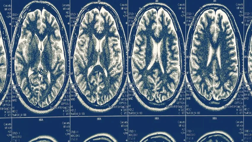
- High-resolution MRI - with special thin-slice sequences (such as 3D CISS/FIESTA and MR angiography) to visualise the nerve and any compressing vessel, and to exclude tumors or multiple sclerosis.
- Response to medicines - a good initial response to carbamazepine supports the diagnosis of trigeminal neuralgia.
- Electrophysiology - in hemifacial spasm, specific EMG findings can support the diagnosis and are used during surgery to confirm adequate decompression.
It is important to note that MRI may sometimes show a vessel near the nerve in people without symptoms, and may not always show a compressing vessel that is found at surgery. Therefore, the clinical picture remains central to decision-making.
Medical Treatment - The First Step
Trigeminal neuralgia is usually treated first with medicines that calm nerve activity. Carbamazepine and its relative oxcarbazepine are the first-choice drugs and are highly effective initially in most patients. Other medicines such as gabapentin, pregabalin, baclofen or lamotrigine may be added. Ordinary painkillers are generally ineffective. Problems arise when the medicines stop working over time, or when the doses needed cause side effects such as drowsiness, dizziness, unsteadiness, confusion, low sodium levels, rashes or liver effects - particularly troublesome in older people. When medicines fail or are poorly tolerated, surgical options should be considered.
For hemifacial spasm, botulinum toxin injections into the affected muscles are the most common non-surgical treatment. They weaken the muscles temporarily and control spasms for about three to four months, after which repeat injections are needed. Many patients do well with this approach, but some find the repeated injections inconvenient, experience side effects such as eyelid drooping, or find the effect diminishing over time. For these patients, MVD offers a potential permanent cure.
Surgical and Procedural Options for Trigeminal Neuralgia
Microvascular decompression (MVD)
Treats the cause by separating the vessel from the nerve. Offers the longest-lasting relief with preservation of facial sensation. Requires a small craniotomy under general anaesthesia.
Percutaneous procedures
Radiofrequency rhizotomy, balloon compression or glycerol injection through a needle in the cheek. Less invasive, quicker recovery, but usually cause some facial numbness and pain may recur sooner.
Stereotactic radiosurgery
Focused radiation to the nerve root without incision. Relief develops over weeks to months; some numbness can occur.
Neurectomy
Cutting peripheral branches - rarely used now due to higher recurrence.
MVD is generally preferred for patients who are fit for surgery and have classical trigeminal neuralgia, especially younger and middle-aged patients, because it offers the best long-term results without deliberately damaging the nerve. Percutaneous procedures and radiosurgery are valuable for older or medically unfit patients, those with multiple sclerosis, or those who prefer to avoid open surgery.
The Microvascular Decompression Procedure
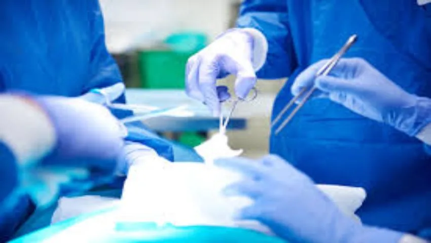
- Anaesthesia and positioning: under general anaesthesia, the patient is positioned on the side with the head secured.
- Monitoring: for hemifacial spasm, facial nerve monitoring and brainstem auditory evoked potentials (to protect hearing) are used; in trigeminal cases, hearing monitoring is often used as well.
- Incision and craniotomy: a small incision behind the ear is followed by a small opening in the skull, about the size of a coin (retrosigmoid keyhole approach).
- Accessing the nerve: under the operating microscope, the dura is opened and cerebrospinal fluid is released so the cerebellum relaxes and can be gently protected, giving access to the cerebellopontine angle.
- Identifying the compression: the nerve is inspected along its length to its root entry zone, and the compressing artery or vein is identified. The endoscope can help visualise hidden areas around the nerve.
- Decompression: the vessel is carefully mobilised away from the nerve, and a small piece of Teflon felt is placed as a cushion between them, or the vessel is transposed and secured away from the nerve.
- Confirmation: in hemifacial spasm, disappearance of the abnormal lateral spread response on monitoring confirms successful decompression.
- Closure: the dura is closed watertight, the bone opening is repaired, and the scalp is closed.
The operation typically takes two to three hours. Most patients spend one night in the intensive care unit for observation and go home within a few days.
Results of Microvascular Decompression
MVD has one of the highest success rates among treatments for trigeminal neuralgia. A large majority of patients with classical trigeminal neuralgia experience immediate or rapid pain relief after surgery, and many remain pain-free for many years without medication. Long-term studies show that a substantial proportion remain free of pain a decade after surgery. Results are best in patients with typical, paroxysmal pain and clear arterial compression; outcomes are less predictable when there is constant background pain or compression by a vein only.
For hemifacial spasm, most patients experience resolution or major improvement of spasms. In some, the twitching stops immediately; in others, it fades gradually over weeks to months as the nerve recovers - so patience is sometimes needed. Recurrence is uncommon after successful decompression.
Risks of MVD
MVD is a delicate operation near the brainstem, but in experienced hands serious complications are uncommon. Possible risks include:
- Hearing loss on the operated side - minimised with auditory monitoring.
- Facial numbness or weakness - usually mild and temporary if it occurs.
- Cerebrospinal fluid leak from the wound - usually treatable.
- Headache, dizziness or imbalance in the first days.
- Infection, bleeding or stroke - rare.
- Recurrence of pain or spasm in a minority of patients over time.
The risks are carefully balanced against the burden of ongoing severe pain or spasm and the side effects of long-term medicines, and are discussed openly with each patient.
Recovery After MVD
Typical recovery
- Hospital stay: usually 3-5 days.
- Medicines: trigeminal neuralgia medicines are usually tapered gradually after surgery under medical supervision.
- Activity: walking from the day after surgery; avoid heavy lifting and straining for about 4-6 weeks.
- Return to work: typically within 2-4 weeks for desk jobs.
- Wound care: keep the incision dry until advised; stitches removed in 10-14 days.
- Follow-up: clinical review to assess relief and taper medicines.
Who Is a Good Candidate for MVD?
- Patients with classical trigeminal neuralgia whose pain is not adequately controlled by medicines, or who cannot tolerate the side effects.
- Patients with hemifacial spasm who wish for a lasting solution rather than repeated botulinum injections, or who no longer respond to them.
- Patients with glossopharyngeal neuralgia refractory to medicines.
- Patients in reasonable general health who can safely undergo general anaesthesia - age alone is not a barrier, and many healthy older adults do very well.
- Ideally, MRI showing a vessel in contact with the nerve, although surgery may still be considered in typical cases without clear imaging findings.
Living With Trigeminal Neuralgia - Practical Tips
While treatment is being optimised, several practical measures can help reduce triggers. Using a soft toothbrush with lukewarm water, eating soft foods at room temperature, drinking through a straw, and protecting the face from wind and cold with a scarf can reduce attacks. Keeping a pain diary helps identify triggers and track the effect of medicines. Taking medicines regularly as prescribed - rather than only when pain strikes - is essential, since they work by preventing attacks. Never stop medicines abruptly. Because severe facial pain can affect nutrition, maintaining adequate calorie and fluid intake is important. Emotional support matters too: the unpredictability of attacks can cause anxiety and depression, and speaking openly with doctors and family helps.
Why Choose Dr. (Prof.) Sumiet Snha for MVD?
- AIIMS-trained neurosurgeon - MBBS, MS, DNB, MCh (AIIMS, New Delhi), FACS - with over 25 years of experience in brain and skull base surgery.
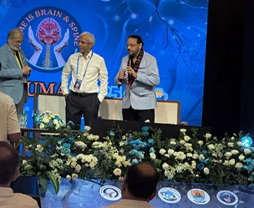
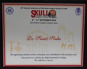
- Former Professor of Neurosurgery, AIIMS, New Delhi, and invited faculty at national skull base surgery meetings.
- Expertise in keyhole retrosigmoid approaches with endoscope assistance for complete visualisation of the nerve.
- Use of intra-operative neuromonitoring to protect hearing and facial nerve function.
- Comprehensive evaluation including high-resolution MRI and discussion of all options - medical, percutaneous, radiosurgical and MVD.
- Advanced neurosurgical infrastructure at Max Super Speciality Hospital, Dwarka.
Cost Factors for MVD Surgery
- Pre-operative imaging with specialised MRI sequences.
- Neuromonitoring and endoscopic equipment.
- Duration of hospital stay, including ICU observation.
- Choice of treatment - MVD versus percutaneous procedures or radiosurgery.
- Insurance coverage.
Trigeminal Neuralgia and Multiple Sclerosis
People with multiple sclerosis (MS) have a higher risk of developing trigeminal neuralgia, and in them the condition may occur at a younger age and sometimes on both sides. In MS, the pain is often caused by a demyelinating plaque in the brainstem affecting the trigeminal pathway rather than by vascular compression, although both may coexist. Medicines remain the first line of treatment, but they may be less well tolerated in people with MS, who often take other medicines and may experience more side effects. When medicines fail, percutaneous procedures and radiosurgery are frequently preferred, while MVD is considered selectively when clear vascular compression is present. Close collaboration with the patient's neurologist ensures coordinated care.
Endoscope-Assisted MVD
The operating microscope provides a straight-line view, which means that some parts of the nerve - particularly the side facing away from the surgeon or the area close to the brainstem - may be difficult to see without retracting the cerebellum. The endoscope, with its angled lenses, can look around corners and illuminate these hidden areas. Using the endoscope during MVD helps the surgeon confirm that all compressing vessels have been identified, check the position of the Teflon cushion, and reduce the need for brain retraction. This combination of microscope and endoscope, which reflects Dr. Snha's broader expertise in endoscopic neurosurgery, supports thorough and gentle decompression.
When Pain Returns After Treatment
Although most patients enjoy long-term relief, pain can return after any treatment for trigeminal neuralgia. After MVD, recurrence may be due to a new vessel coming into contact with the nerve, slippage of the cushion, scarring or progression of nerve damage. After percutaneous procedures or radiosurgery, pain recurrence is more common over time as the nerve partially recovers. Recurrent pain is not the end of the road: medicines often work again, and repeat or alternative procedures can be considered. A careful review of the pain pattern, a fresh MRI and a discussion of all options help choose the best next step.
Preparing for MVD Surgery
Once the decision for microvascular decompression has been made, a few preparatory steps help ensure that the operation is as safe as possible and that recovery is smooth. Each patient receives individual instructions, but the general sequence is as follows.
- Review of imaging: the high-resolution MRI is studied carefully to plan the approach and anticipate the likely compressing vessel.
- Fitness for anaesthesia: blood tests, ECG and, where needed, heart or lung assessment. Blood pressure and diabetes should be well controlled.
- Blood thinners: aspirin, clopidogrel and anticoagulants usually need to be paused before surgery, but only after discussion with the doctor who prescribed them.
- Trigeminal neuralgia medicines: these are continued up to the day of surgery and should never be stopped suddenly beforehand.
- Hearing test: a baseline audiogram is often performed, since the nerve that controls hearing lies close to the operative area.
- Consent discussion: the expected benefits, risks and alternatives are reviewed again so that every question is answered before the day of surgery.
Patients with hemifacial spasm who receive botulinum toxin injections should mention the date of their last injection, because the remaining effect of the toxin can influence the assessment of spasm before and after surgery.
Warning Signs After Going Home
Most patients recover without difficulty after MVD, but it is important to know which symptoms need prompt attention. Some tiredness, mild headache, discomfort at the wound and a slight feeling of imbalance are common in the first days and gradually settle.
Contact the team promptly if you notice
- Clear, watery fluid dripping from the nose or from the wound, especially when bending forward - this may be a cerebrospinal fluid leak.
- Fever, neck stiffness or a severe, worsening headache.
- Redness, swelling or discharge at the incision.
- New weakness of the face, difficulty closing the eye, or new hearing loss.
- Persistent vomiting, confusion or increasing drowsiness.
Most such problems can be managed simply when recognised early. Patients should also avoid blowing the nose forcefully and straining for the first few weeks, and follow advice on bathing and wound care until the incision has healed. Any change in medicines, particularly the tapering of carbamazepine or oxcarbazepine, should be done only as advised at follow-up.
Travelling to Delhi for MVD
Because microvascular decompression is a specialised operation, many patients travel to Delhi from other cities and states, and some from abroad. Careful planning helps reduce the burden of travel. Patients can send their MRI images, a description of their pain or spasm and a list of medicines tried by WhatsApp or e-mail for a preliminary opinion. This allows the team to judge whether MVD is likely to be suitable and whether any additional imaging is needed - for example, specialised thin-slice sequences that were not included in the original scan.
Max Super Speciality Hospital, Dwarka, is close to Indira Gandhi International Airport, which is convenient for patients arriving by air. The likely duration of the hospital stay is explained beforehand, and patients are advised on when it is appropriate to travel home after surgery and by what means. Follow-up after returning home - including the gradual reduction of medicines - can often be guided by video consultation, with the local doctor kept informed through a detailed discharge summary.
Questions to Ask Before Deciding on MVD
Choosing between continuing medicines, MVD, a percutaneous procedure or radiosurgery is a personal decision. It depends on the severity of symptoms, general health, age, the MRI findings and each patient's own priorities. The following questions can help structure the discussion with your neurosurgeon.
| Question | What the answer tells you |
|---|---|
| Is my pain typical of classical trigeminal neuralgia? | Typical, paroxysmal pain is the pattern most likely to respond well to MVD. |
| Does my MRI show a vessel touching the nerve? | A clear arterial contact supports the case for decompression, though it is not the only factor. |
| What are my alternatives, and how do they compare? | Helps weigh long-term relief against the invasiveness of each option. |
| How will my age and health affect the risks? | General fitness, rather than age alone, guides the choice. |
| What will recovery involve for me? | Allows planning of work, travel and family support. |
| What happens if symptoms come back? | Shows that there is a plan beyond the first treatment. |
At the consultation, Dr. Snha reviews each of these points with the patient and family, going through the MRI images on screen. There is no pressure to decide immediately - many patients prefer to take time, discuss with relatives and return with further questions, and this is encouraged.
It can also help to keep a simple record of your symptoms in the weeks before the consultation: how often attacks occur, what triggers them, how long they last, which medicines and doses you have tried, and any side effects you have experienced. For hemifacial spasm, a short video of the spasms recorded on a phone can be very useful, since the twitching may not appear during the appointment itself. This information allows a more accurate assessment and a better-informed decision about whether MVD is the right choice.
Frequently Asked Questions
What causes trigeminal neuralgia?
In most cases, a blood vessel pressing on the trigeminal nerve where it leaves the brainstem damages its insulation, causing abnormal pain signals. Less commonly, it is caused by multiple sclerosis or a tumor.
Is MVD a permanent cure?
MVD provides long-lasting relief in most patients with classical trigeminal neuralgia, with many remaining pain-free for many years. A minority may experience recurrence over time.
Will I have facial numbness after MVD?
MVD aims to preserve normal facial sensation. Numbness is uncommon and usually mild, unlike destructive procedures, which intentionally cause some numbness.
Can hemifacial spasm be cured?
Yes. MVD cures or significantly improves hemifacial spasm in most patients, although improvement may take weeks to months in some.
Am I too old for MVD?
Age alone does not rule out MVD. Healthy older adults often do very well. For those with significant medical problems, less invasive options are available.
How long is the hospital stay?
Usually 3 to 5 days, including a night of ICU observation after surgery.
Can trigeminal neuralgia be mistaken for a dental problem?
Yes. Many patients initially consult a dentist. If tooth or jaw pain is sharp, electric and triggered by touch or chewing, trigeminal neuralgia should be considered.
Relief Is Possible
If facial pain or spasm is controlling your life, you do not have to suffer indefinitely. Book a consultation with Dr. (Prof.) Sumiet Snha at Max Hospital, Dwarka, New Delhi, to explore whether microvascular decompression or another treatment is right for you. Related pages: acoustic neuroma, chronic nerve pain and spasticity treatment.