Every nerve in the body - from those inside the skull to those in the fingertips - is wrapped in protective sheaths. The most important of these are formed by Schwann cells, which wrap around nerve fibres like insulation around electrical wires. When these sheath cells grow abnormally, they form nerve sheath tumors. Because nerves run throughout the body, nerve sheath tumors can appear in very different places: inside the skull on the nerves of hearing or facial sensation, within the spinal canal on the nerve roots, straddling the spine and chest in a dumbbell shape, or along the nerves of the arms and legs.
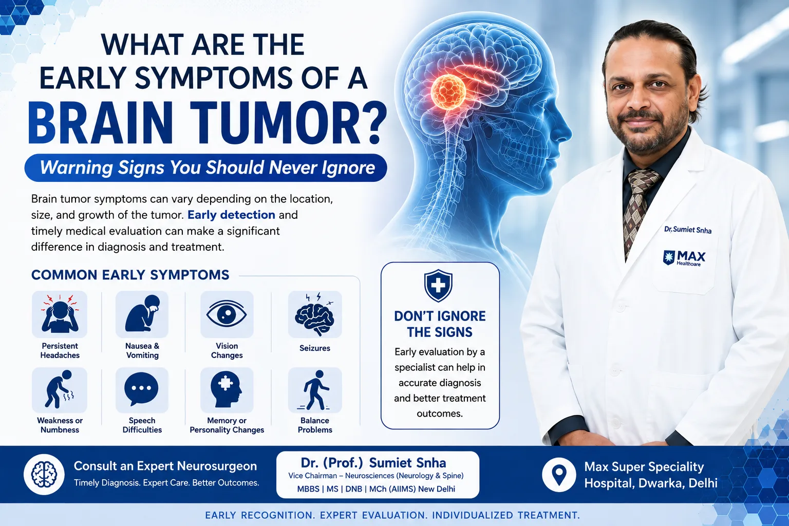
Although these tumors share a common origin, the challenges they pose depend greatly on their location. A schwannoma on the ulnar nerve at the elbow and a schwannoma on a spinal nerve root pressing on the spinal cord are the same type of tumor, but they require different evaluation, surgical approaches and expertise. This guide by Dr. (Prof.) Sumiet Snha - a neurosurgeon with specialist expertise in brain, spine and peripheral nerve surgery, Founder Secretary of the Indian Society of Peripheral Nerve Surgery and former Professor of Neurosurgery at AIIMS, New Delhi - explains nerve sheath tumors by location, so patients understand their specific situation. For a general overview of lumps on limb nerves, see our nerve tumor page.
The Nerve Sheath - Understanding the Anatomy
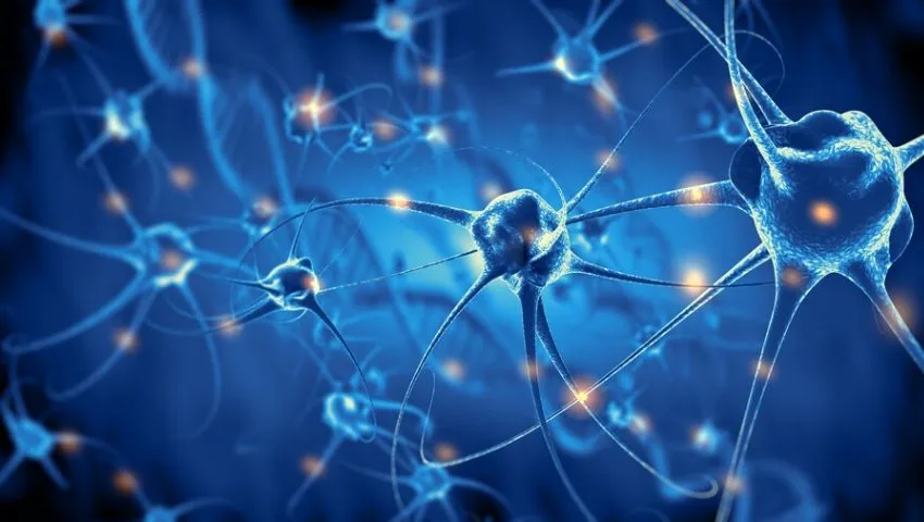
A peripheral nerve has three layers of connective tissue. The endoneurium surrounds individual nerve fibres, the perineurium wraps groups of fibres into fascicles, and the epineurium encloses the whole nerve. Schwann cells lie along each nerve fibre, producing myelin that speeds up nerve signals. Nerve sheath tumors originate mainly from Schwann cells (schwannomas), from a mixture of Schwann cells, fibroblasts and other cells (neurofibromas), or rarely from perineurial cells (perineuriomas). Central nervous system tissue - the brain and spinal cord - uses different cells (oligodendrocytes) for myelination, which is why nerve sheath tumors arise on nerves after they leave the brain or spinal cord, including the cranial nerves and spinal nerve roots.
Types of Nerve Sheath Tumors
Schwannoma
Encapsulated, eccentric growth pushing fibres aside; usually removable completely with nerve preservation. The most common type.
Neurofibroma
Unencapsulated, grows among nerve fibres; can be localised, diffuse or plexiform; associated with NF1.
Perineurioma
Rare, benign tumor of perineurial cells, often causing gradual weakness of a single nerve in young people.
Hybrid tumors
Show features of more than one type, especially in genetic syndromes.
Malignant peripheral nerve sheath tumor
A rare, aggressive sarcoma requiring wide excision and oncological treatment.
Melanotic schwannoma
An uncommon variant containing pigment, requiring careful follow-up.
Nerve Sheath Tumors Inside the Skull
Among the twelve pairs of cranial nerves, the vestibular (balance) nerve is by far the most frequently affected, producing the vestibular schwannoma or acoustic neuroma, which causes hearing loss, tinnitus and imbalance - discussed in detail on our acoustic neuroma page. Less common cranial schwannomas include:
- Trigeminal schwannoma - arising from the nerve of facial sensation, causing facial numbness, pain or weakness of chewing muscles. It may straddle the middle and posterior cranial fossae, requiring skull base approaches.
- Facial nerve schwannoma - causing facial twitching or weakness and hearing changes; management focuses on preserving facial function.
- Jugular foramen schwannomas - arising from the nerves controlling swallowing, voice and shoulder movement.
- Hypoglossal schwannoma - causing weakness and wasting of one side of the tongue.
These tumors are treated with skull base microsurgery, radiosurgery or observation depending on size, growth and symptoms, with careful protection of neighbouring nerves and the brainstem.
Spinal Nerve Sheath Tumors
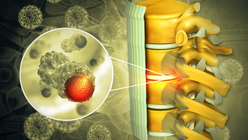
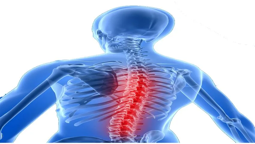
Schwannomas and neurofibromas are among the most common tumors found inside the spinal canal. They usually arise from the sensory (dorsal) nerve roots and lie inside the dura but outside the spinal cord - the "intradural-extramedullary" compartment. As they grow slowly, they compress the spinal cord or the cauda equina (the bundle of nerve roots in the lower back).
Symptoms of spinal nerve sheath tumors
- Radicular pain - pain along the course of the affected nerve root, such as a band of pain around the chest or abdomen (in the thoracic spine), arm pain (in the neck) or sciatica (in the lower back). This pain is often worse at night or on lying down.
- Spinal cord compression symptoms - stiffness and heaviness of the legs, unsteady walking, numbness spreading up from the feet and, later, bladder disturbances.
- Cauda equina symptoms - leg pain, numbness, weakness and bladder or bowel problems in lumbar tumors.
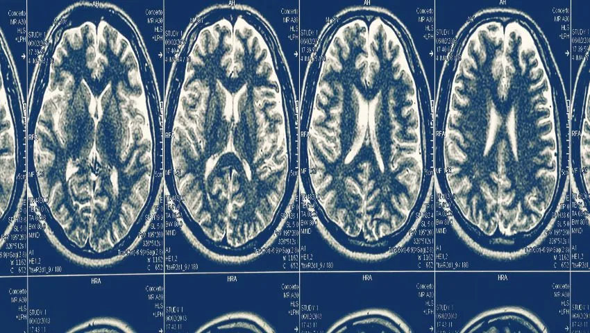
Because these symptoms develop slowly and resemble a slip disc or arthritis, spinal schwannomas are often diagnosed late. An MRI of the spine with contrast clearly shows them.
Treatment of spinal schwannomas
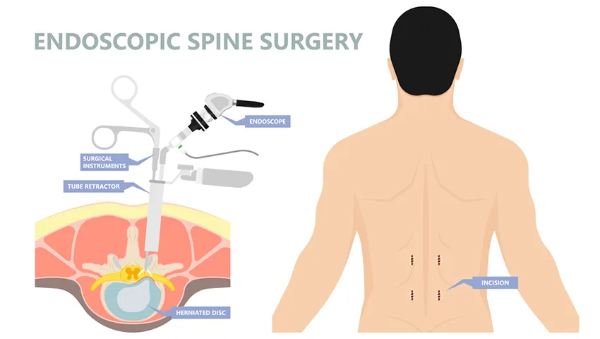
Symptomatic spinal schwannomas are removed surgically. Through a laminectomy or, in many cases, a minimally invasive hemilaminectomy with tubular retractors and the operating microscope or endoscope, the dura is opened and the tumor is separated from the spinal cord and adjacent nerve roots. The small rootlet from which it arises - usually a non-functioning sensory rootlet - is divided. Neuromonitoring protects spinal cord and nerve function. Complete removal usually results in a cure, and patients often experience rapid relief of pain and progressive improvement in walking and sensation. More detail on spinal tumor surgery is available on our spinal tumor page.
Dumbbell Tumors - Crossing Boundaries
Some nerve sheath tumors grow along the nerve root through the intervertebral foramen - the bony opening where the nerve leaves the spine - and expand on both sides, creating a dumbbell shape with one part inside the spinal canal and another in the neck, chest (paraspinal or mediastinal) or abdomen (retroperitoneal). These tumors widen the foramen, sometimes eroding bone, and can be surprisingly large before causing symptoms.
Removing dumbbell tumors requires planning to address both the intraspinal and extraspinal components safely. Depending on size and location, surgery may be performed through a single posterior approach with removal of part of the facet joint, a combined posterior and anterior approach (for example, with a thoracic or abdominal surgeon), or a staged approach. When significant bone is removed, the spine may need to be stabilised with screws. In the neck, the vertebral artery lies close to the foramen and must be protected. A surgeon with expertise in both spine and peripheral nerve surgery is well placed to manage these complex tumors.
Nerve Sheath Tumors of the Brachial and Lumbosacral Plexus
Tumors of the brachial plexus - the nerve network in the neck and shoulder - may present as a lump above the collarbone, pain or tingling in the arm, or weakness. They lie in a region crowded with major blood vessels, the lung apex and important nerves. Surgical exposure of the plexus with identification of all its components, nerve stimulation and meticulous dissection allows safe removal. Similarly, tumors of the lumbosacral plexus in the pelvis may cause leg pain and weakness and require specialised abdominal or retroperitoneal approaches, sometimes in collaboration with general or gynaecological surgeons.
Schwannoma vs Neurofibroma - Key Differences
Growth pattern
Schwannoma: eccentric, pushes fibres aside. Neurofibroma: fusiform, fibres run through the tumor.
Capsule
Schwannoma: well-defined capsule. Neurofibroma: usually no true capsule.
Surgical removal
Schwannoma: usually complete with nerve preservation. Neurofibroma: often partial to protect function.
Association
Schwannoma: sporadic, NF2, schwannomatosis. Neurofibroma: sporadic or NF1.
Malignant potential
Schwannoma: extremely rare. Neurofibroma (plexiform): small lifetime risk of MPNST.
Pain
Both can cause pain; schwannomas often cause tingling on pressure; painful multiple tumors suggest schwannomatosis.
What the Pathology Report Tells You
After surgery, the removed tissue is examined by a pathologist. The report confirms the tumor type using the appearance of the cells and special stains - for example, schwannomas typically show strong, diffuse staining for a protein called S100 and show characteristic cellular patterns (Antoni A and Antoni B areas, and Verocay bodies). Neurofibromas show a mixture of cell types. The report also comments on features such as cell division rate and necrosis that could suggest malignancy. In most cases the report confirms a benign tumor, and no further treatment beyond follow-up is needed. If unusual features are present, additional tests and a discussion about further treatment follow.
The Patient Journey - From Diagnosis to Recovery
- First consultation: history, examination (including checking for skin signs of neurofibromatosis) and review of existing scans.
- Imaging: MRI with contrast of the relevant region; ultrasound for limb tumors; spine and brain MRI if a genetic condition is suspected.
- Nerve studies: to document baseline function.
- Decision: observation versus surgery (or radiosurgery for some cranial tumors), discussed in detail with the patient.
- Surgery: microsurgical removal with nerve monitoring, using minimally invasive approaches where suitable.
- Hospital stay: from a day for limb tumors to several days for spinal or skull base tumors.
- Recovery: gradual return to activities over weeks; physiotherapy if needed.
- Follow-up: clinical review, pathology discussion and imaging as appropriate.
Possible Risks of Surgery
For peripheral schwannomas, the main risks are temporary numbness or tingling, infection and, uncommonly, lasting weakness or sensory loss. For spinal tumors, additional risks include cerebrospinal fluid leak and, rarely, worsening of spinal cord function. For cranial nerve tumors, risks relate to nearby nerves - for example, facial weakness or swallowing difficulties. These risks are minimised through careful planning, microsurgical technique and neuromonitoring, and are discussed in detail before surgery.
Radiosurgery for Nerve Sheath Tumors
Stereotactic radiosurgery is an established option for some cranial nerve schwannomas, particularly small to medium vestibular schwannomas, and occasionally for spinal schwannomas in patients who are not suitable for surgery or who have residual or recurrent tumor. Radiosurgery aims to stop tumor growth rather than remove it. It is generally not used for peripheral limb schwannomas, which are readily and safely removed surgically.
Schwannomatosis - When Pain Is the Main Problem
Schwannomatosis is a condition in which multiple schwannomas develop on peripheral and spinal nerves, without the bilateral vestibular schwannomas typical of NF2. The dominant symptom is often chronic pain, which may be widespread and difficult to control. Some cases are linked to changes in specific genes (such as SMARCB1 or LZTR1) and can run in families. Management combines targeted surgical removal of the tumors causing the most pain or neurological problems with a comprehensive pain management plan, including medicines for neuropathic pain, physiotherapy and psychological support. Whole-body MRI may be used to map tumors. Because new tumors can develop over time, long-term follow-up is needed.
Why Choose Dr. (Prof.) Sumiet Snha?
- A rare combination of expertise in brain, spine and peripheral nerve surgery - covering nerve sheath tumors in every location.
- Founder Secretary, Indian Society of Peripheral Nerve Surgery, with more than 250 complex nerve operations.
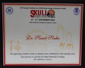
- Former Professor of Neurosurgery, AIIMS, New Delhi, and invited faculty at national skull base and nerve surgery meetings.
- Minimally invasive spinal tumor removal, dumbbell tumor surgery, plexus surgery and skull base microsurgery.
- Neuromonitoring, navigation and multidisciplinary oncology and genetics support at Max Super Speciality Hospital, Dwarka.
Cost Considerations
- Location of the tumor - limb, plexus, spine, skull base.
- Surgical approach and need for stabilisation or combined approaches.
- Neuromonitoring and imaging.
- Hospital stay and rehabilitation.
- Insurance coverage.
Nerve Sheath Tumors in Children and Young Adults
Nerve sheath tumors in children most often occur in the context of neurofibromatosis type 1, where plexiform neurofibromas may be present from early childhood and can grow during periods of rapid growth such as puberty. They may cause visible swelling, asymmetry of a limb or the face, pain, or pressure on important structures such as the airway or spinal cord. Management is multidisciplinary and increasingly includes targeted medical therapy with MEK inhibitors, which can shrink plexiform neurofibromas in many children, reducing pain and improving function. Surgery remains important for tumors causing compression, for diagnostic uncertainty, or when there are concerns about malignant change. Solitary schwannomas are uncommon in children and warrant evaluation for NF2 or schwannomatosis.
Returning to Normal Life After Surgery
After removal of a peripheral nerve sheath tumor, most people return to office work within one to two weeks and to more physical work within a few weeks, depending on the location of the surgery. After spinal tumor removal, patients typically walk the next day, go home within a few days and gradually increase activity over four to six weeks, avoiding heavy lifting initially. Physiotherapy helps restore strength and balance where the tumor had caused weakness or walking difficulty. Numbness in a small patch of skin corresponding to the sensory rootlet of origin is common and usually not bothersome. Most patients are delighted to find that the pain which may have troubled them for months or years has resolved.
Common Misdiagnoses
Nerve sheath tumors are frequently mistaken for other conditions, delaying the right treatment. Limb schwannomas are often thought to be ganglion cysts, lipomas or enlarged lymph nodes. Spinal schwannomas causing sciatica or arm pain are often attributed to disc disease, and patients may receive prolonged treatment for a "slip disc" before an MRI with contrast reveals the tumor. Thoracic spinal schwannomas causing band-like chest or abdominal pain may be investigated for heart, lung or gastrointestinal causes. Awareness of these patterns and appropriate imaging - particularly contrast MRI when pain is persistent, worse at night, or accompanied by neurological signs - leads to earlier diagnosis.
Neuromonitoring - Protecting Nerve Function During Surgery
The central challenge in removing a nerve sheath tumor is taking out the abnormal tissue while leaving the working nerve fibres intact. Under the operating microscope, tumor and nerve can look similar, and the fascicles that pass over a schwannoma's surface may be stretched thin. Intra-operative neurophysiological monitoring gives the surgeon real-time information about nerve function, so that decisions are based on how the nerve behaves electrically rather than on appearance alone. It is a key safety measure in spinal, plexus and skull base tumor surgery, and is used selectively for limb tumors.
Techniques commonly used
- Direct nerve stimulation - a fine probe delivers a tiny electrical current to fascicles on the tumor surface. If a muscle twitches, the fascicle is functional and is protected; if not, it may be a non-functioning fascicle entering the tumor that can safely be divided.
- Free-running electromyography (EMG) - recording electrodes in the relevant muscles detect irritation of a nerve during dissection and alert the surgical team.
- Motor evoked potentials (MEPs) - used in spinal tumor surgery to confirm that the pathways controlling leg and arm movement remain intact while the tumor is separated from the spinal cord.
- Somatosensory evoked potentials (SSEPs) - track the sensory pathways through the spinal cord or plexus.
- Nerve action potentials - in selected cases, measure conduction across a segment of nerve to guide decisions about reconstruction.
Monitoring requires coordination between the surgeon, anaesthetist and neurophysiologist; for example, certain muscle-relaxing drugs are avoided so that responses can be recorded. Although monitoring cannot remove every risk, it helps the surgeon achieve the fullest safe removal and reduces the chance of unexpected weakness after surgery.
Nerve Sheath Tumors Found by Chance
With the increasing use of MRI and CT scans, many nerve sheath tumors are now discovered incidentally - for example, a small schwannoma seen on a lumbar spine MRI done for back pain, or a paraspinal mass noticed on a chest CT performed for a cough. Finding an unexpected "tumor" on a report is understandably alarming, but most such lesions are small, benign and slow-growing. The first step is to confirm, with dedicated contrast-enhanced imaging, that the appearance is typical of a nerve sheath tumor and to examine the patient carefully for any subtle symptoms or neurological signs that may have been overlooked.
Small, symptom-free tumors with typical imaging features can often be safely observed with a repeat scan after several months, then at longer intervals if they remain unchanged. Surgery is considered if the tumor grows, begins to compress the spinal cord or nerve roots, causes symptoms, or has features that raise doubt about the diagnosis. The decision takes into account the tumor's location, the patient's age and general health, and their own preferences after a clear discussion of the options.
Warning Signs That Need Prompt Review
Most nerve sheath tumors behave predictably and change slowly over years. A small number of situations, however, should prompt an early specialist review rather than waiting for a routine appointment. These signs do not necessarily mean that a tumor is malignant or dangerous, but they indicate that the situation has changed and needs fresh assessment, often with repeat imaging.
Seek early review if you notice
Rapid enlargement of a known lump over weeks to a few months; new, persistent pain that is severe or wakes you at night; new weakness, numbness or wasting of muscles in the limb supplied by the nerve; a lump that becomes hard and fixed; difficulty walking, stiffness of the legs or new bladder or bowel changes in someone with a spinal tumor; or, in people with NF1, a plexiform neurofibroma that suddenly becomes painful or starts to grow.
In people with neurofibromatosis type 1, where the risk of malignant change is higher than in the general population, a change in a plexiform neurofibroma is investigated promptly, sometimes with specialised scans such as PET-CT to identify areas of increased activity that may need biopsy. Early recognition allows the best chance of effective treatment.
Long-Term Follow-Up After Treatment
For most people with a single, completely removed schwannoma, follow-up is straightforward: a clinical review to check wound healing and nerve function, discussion of the pathology report and, for spinal tumors, a follow-up MRI to confirm complete removal. If that scan is clear and the patient is well, further imaging may be infrequent or unnecessary. Recurrence after complete removal of a benign schwannoma is uncommon.
Follow-up is more structured when a tumor has been partially removed to protect nerve function, when a neurofibroma has been debulked, or when the patient has NF1, NF2 or schwannomatosis. In these situations, periodic clinical examination and imaging help detect growth of residual tumor or new tumors early, when treatment options are simplest. For people with genetic conditions, follow-up often involves several specialists - including neurologists, geneticists, ophthalmologists and audiologists - and Dr. Snha coordinates the neurosurgical aspects of this care at Max Super Speciality Hospital, Dwarka.
Questions Worth Asking Before Surgery
- How confident are we about the diagnosis from the imaging, and is a biopsy needed?
- Can the tumor be removed completely, or is partial removal safer for this nerve?
- Which nerve functions are at risk, and how will they be protected during surgery?
- Will a minimally invasive approach be possible in my case?
- How long will I stay in hospital, and when can I return to work and exercise?
- What follow-up will I need, and should I be tested for a genetic condition?
Patients are encouraged to bring these and their own questions to the consultation. Reports can also be shared in advance on WhatsApp at +91-8448877746 for a preliminary opinion, or you can book an appointment directly.
The Multidisciplinary Team Behind Nerve Sheath Tumor Care
Although the operation itself is performed by the neurosurgeon, safe and complete care for a nerve sheath tumor relies on a wider team. Each specialist contributes a different piece of the picture, from the first scan to the final follow-up visit. This team approach is particularly important for tumors in complex locations, for suspected malignant tumors and for people with neurofibromatosis or schwannomatosis, where decisions often involve balancing several competing considerations.
- Neuroradiologist - interprets MRI and CT scans, helps distinguish nerve sheath tumors from other masses and plans image-guided biopsy when needed.
- Neurophysiologist - performs nerve conduction studies before surgery and runs intra-operative monitoring.
- Anaesthetist - tailors anaesthesia to allow reliable nerve monitoring and plans pain relief afterwards.
- Neuropathologist - confirms the diagnosis and looks for any features of concern.
- Thoracic, abdominal or head and neck surgeons - join for dumbbell, mediastinal, pelvic or neck tumors that need combined approaches.
- Oncologist and radiation specialist - involved for malignant tumors or when radiosurgery is considered.
- Geneticist - advises on testing and family implications when a syndrome is suspected.
- Physiotherapist and pain specialist - support recovery of strength, balance and comfort after surgery.
At Max Super Speciality Hospital, Dwarka, these specialists are available under one roof, which allows complex cases to be discussed jointly and a single, coordinated plan to be presented to the patient and family.
Frequently Asked Questions
What is a nerve sheath tumor?
It is a tumor arising from the protective sheath cells surrounding nerves, most commonly a schwannoma or neurofibroma. Most are benign.
Can a spinal schwannoma be removed with keyhole surgery?
Many spinal schwannomas can be removed through minimally invasive approaches with tubular retractors and the microscope or endoscope, depending on size and location.
What is a dumbbell tumor?
A nerve sheath tumor that grows through the opening where a nerve leaves the spine, with parts inside and outside the spinal canal. It may need combined or staged surgery.
Do nerve sheath tumors turn into cancer?
Schwannomas very rarely become malignant. Plexiform neurofibromas in NF1 carry a small lifetime risk of malignant change, so they are monitored closely.
Will I need radiation after surgery?
Not usually for benign tumors that are completely removed. Radiosurgery may be used for some cranial or residual tumors.
Can multiple nerve tumors be a sign of a genetic condition?
Yes. Multiple nerve sheath tumors may indicate NF1, NF2 or schwannomatosis. Genetic evaluation and counselling may be recommended.
How long does it take to recover from spinal schwannoma surgery?
Many patients walk the day after surgery, go home within a few days and return to light activities within a few weeks.
Expert Care for Nerve Sheath Tumors Anywhere in the Body
Whether your tumor is on a limb nerve, a spinal nerve root or a cranial nerve, an experienced specialist can guide you to the safest treatment. Book a consultation with Dr. (Prof.) Sumiet Snha at Max Hospital, Dwarka, New Delhi. Related pages: nerve tumors, spinal tumors and acoustic neuroma.