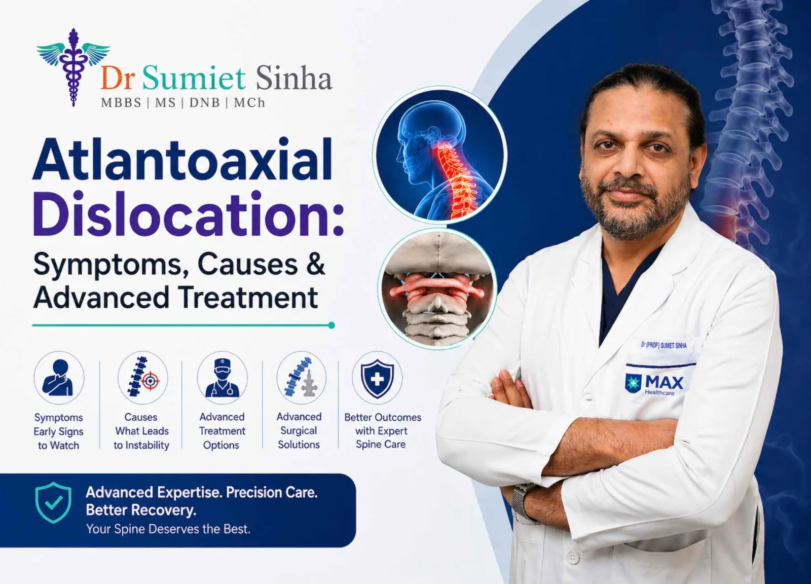
The junction between the skull and the top of the neck - the craniovertebral junction (CVJ) - is one of the most complex and critical regions of the human body. Here, the brainstem continues into the spinal cord, vital arteries wind their way to the brain, and the head rotates and nods on two unique vertebrae: the atlas (C1) and the axis (C2). When the connection between these two bones becomes loose or displaced, the condition is called atlanto-axial dislocation (AAD) or atlanto-axial instability. Because the spinal cord at this level controls breathing, movement of all four limbs and sensation of the entire body, AAD can have serious consequences if not treated correctly.
India has a comparatively high number of patients with congenital CVJ anomalies, and AAD is a condition that Indian neurosurgical centres - AIIMS, New Delhi in particular - have extensive experience in treating. Dr. (Prof.) Sumiet Snha, former Professor of Neurosurgery at AIIMS and Principal Director - Neurosurgery & Spine Surgery at Max Hospital, Dwarka, New Delhi, treats children and adults with congenital, traumatic and inflammatory AAD using modern fixation techniques guided by navigation and neuromonitoring.
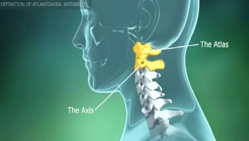
Understanding the Atlas and Axis
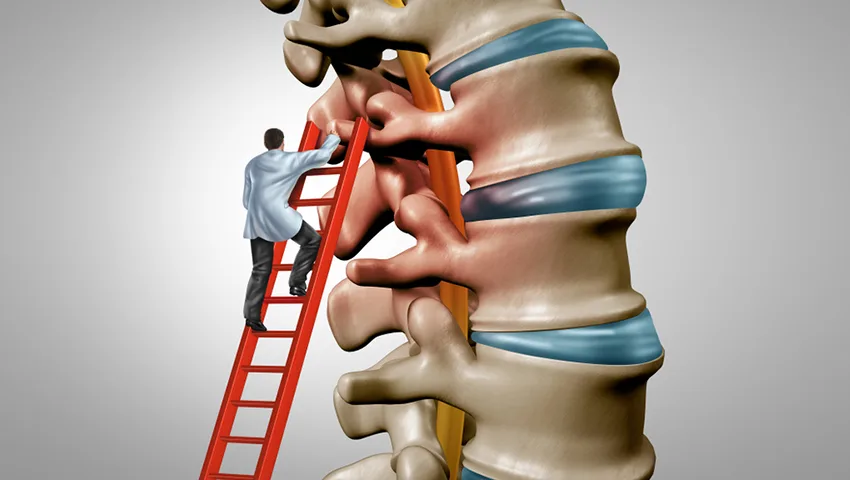
Most vertebrae in the spine look broadly similar - a round body in front and a bony arch behind. The top two are different. The atlas (C1) is a ring of bone without a body, supporting the skull like the mythological Atlas supporting the world. The axis (C2) has a tooth-like projection called the odontoid process or dens, which projects upward into the front of the atlas ring. The dens is held snugly against the atlas by the strong transverse ligament, forming a pivot joint that allows about half of all head rotation.
When the transverse ligament is torn or lax, the dens is fractured or malformed, or the joints between C1 and C2 are abnormal, the atlas can slide forward (or sometimes backward or rotate) on the axis. This reduces the space available for the spinal cord, which may be pinched between the dens in front and the arch of the atlas behind.
What Is Atlanto-Axial Dislocation?
Atlanto-axial dislocation refers to loss of the normal alignment and stability between C1 and C2. It can be:
Reducible AAD
The displacement corrects (reduces) when the neck is extended or placed in traction. Posterior C1-C2 fixation is usually sufficient.
Irreducible AAD
The displacement does not correct with position or traction, often due to long-standing deformity. Special techniques are needed to realign and decompress.
Rotatory subluxation
C1 is rotated and locked on C2, often in children after infection or minor trauma, causing a tilted head ("cock-robin" posture).
AAD with basilar invagination
The dens projects upward into the skull base, compressing the brainstem - often associated with Chiari malformation or syringomyelia.
Causes of Atlanto-Axial Dislocation
Congenital anomalies
Many patients are born with abnormalities of the craniovertebral junction: an os odontoideum (a separate, unfused dens), fusion of the atlas with the skull (atlanto-occipital assimilation), abnormal C1-C2 joints, a short neck with fused vertebrae (Klippel-Feil syndrome) or basilar invagination. These may remain silent for years and first cause symptoms in adolescence or adulthood, sometimes after a minor fall or neck strain.
Trauma
Road traffic accidents, falls from height and sports injuries can fracture the dens or tear the transverse ligament. Odontoid fractures are among the most common cervical spine fractures in the elderly, often after a simple fall at home.
Inflammatory conditions
Rheumatoid arthritis can erode the ligaments and joints around the dens, gradually leading to instability. Ankylosing spondylitis and other inflammatory conditions can also involve this region.
Syndromes
Children with Down syndrome, Morquio syndrome, skeletal dysplasias and some connective tissue disorders have a higher risk of ligament laxity and AAD.
Infections and tumours
Throat or neck infections (Grisel syndrome in children), tuberculosis of the CVJ and tumours can destroy the supporting structures and cause instability.
Congenital Craniovertebral Junction Anomalies in Detail
Os odontoideum
In os odontoideum, the tip of the dens exists as a separate, rounded ossicle that is not firmly joined to the body of C2. Whether it results from an unrecognised childhood injury or a developmental failure, the effect is the same: the ligaments no longer hold C1 securely to C2, and the atlas can glide forwards or backwards with neck movement. Some people discover it only on an X-ray taken for another reason; others present with myelopathy or even sudden weakness after a fall. Because instability can be unpredictable, symptomatic os odontoideum is usually treated with C1-C2 fixation.
Atlanto-occipital assimilation
Here the atlas is partially or completely fused with the base of the skull. The joints below then bear abnormal stress and gradually become lax, allowing the dens to move backwards and upwards. Assimilation is frequently combined with basilar invagination and Chiari malformation, and it changes the surgical plan because a conventional C1 screw may not be possible.
Basilar invagination
In basilar invagination the tip of the dens rises above its normal position and indents the foramen magnum, pressing on the brainstem from the front. Patients may have a short neck, a low hairline, restricted neck movement, headaches, swallowing difficulties, nystagmus or weakness. Two broad groups are recognised: one with C1-C2 joint instability, where joint distraction and fixation can bring the dens down; and another without instability, where decompression at the skull base may be the priority.
Klippel-Feil syndrome
This condition involves congenital fusion of two or more cervical vertebrae. The fused segments place extra stress on the remaining mobile levels, which may become unstable or degenerate early. Associated anomalies of the heart, kidneys or hearing are sometimes present and are assessed before surgery.
Traumatic Injuries of C1 and C2
High-energy accidents in young adults and simple falls in the elderly are the two main scenarios for upper cervical injuries.
Jefferson (C1 ring) fracture
A burst fracture of the atlas ring from axial load, as in diving into shallow water. Stability depends on whether the transverse ligament is intact.
Odontoid fractures (Types I-III)
Type II fractures at the base of the dens have the poorest natural healing and often need screw fixation or C1-C2 fusion.
Hangman's fracture
A fracture through the bony bridges (pars) of C2, sometimes with forward slippage of C2 on C3. Many heal in a collar; displaced ones need fixation.
Transverse ligament rupture
A purely ligamentous injury that will not heal reliably on its own, usually requiring surgical stabilisation.
Every patient with a significant head or neck injury should be assessed with appropriate imaging before the neck is moved freely. Proper immobilisation at the scene and in the emergency department - principles taught in ATLS courses, for which Dr. Snha is certified faculty - prevents secondary spinal cord injury.
Rheumatoid Arthritis and the Upper Neck
In long-standing rheumatoid arthritis, inflammatory tissue (pannus) erodes the ligaments and bone around the dens. This can lead to forward slippage of C1 on C2, vertical settling of the skull on the spine and compression of the brainstem. Symptoms may be masked by joint pain elsewhere, so patients with rheumatoid arthritis who develop neck pain, occipital headache, tingling in the hands or difficulty walking should have their upper cervical spine evaluated. Anaesthesia teams also screen these patients before any surgery, because intubation can stress an unstable neck. Stabilisation surgery, when needed, is coordinated with the rheumatologist to optimise medicines and bone health.
Symptoms of Atlanto-Axial Dislocation
Symptoms range from none at all to severe neurological deficit, and may develop gradually or suddenly after a trivial injury:
- Neck pain, especially at the back of the upper neck and base of the skull.
- Restricted or painful neck movement, head tilt or torticollis.
- Occipital headache radiating to the back of the head.
- Weakness in the arms and/or legs, clumsiness of the hands.
- Numbness and tingling in the limbs or body.
- Unsteady gait, frequent falls, stiffness of the legs (spasticity).
- Brainstem symptoms - difficulty swallowing, change in voice, dizziness, double vision or episodes of breathlessness, particularly when basilar invagination is present.
- Transient symptoms such as tingling or "electric shock" sensations on bending the neck (Lhermitte's sign).
- Bladder and bowel disturbance in advanced cases.
An important safety warning
People with known AAD or suspected instability should avoid neck manipulation, forceful massage, "cracking" of the neck, and sudden jerky movements, as these can cause sudden spinal cord injury. Any worsening weakness, breathing difficulty or new bladder problem requires immediate medical attention.
How AAD Is Diagnosed
- Clinical examination - neck posture, range of movement (performed cautiously), neurological assessment of all four limbs and cranial nerves.
- Dynamic X-rays - lateral views of the neck in flexion and extension, performed under supervision, to measure the atlanto-dental interval and assess reducibility.
- CT scan with 3D reconstruction - essential to understand the bony anatomy, joint orientation, os odontoideum and fractures, and to plan screw trajectories.
- CT angiography - to map the course of the vertebral arteries, which vary considerably in CVJ anomalies and must be protected during surgery.
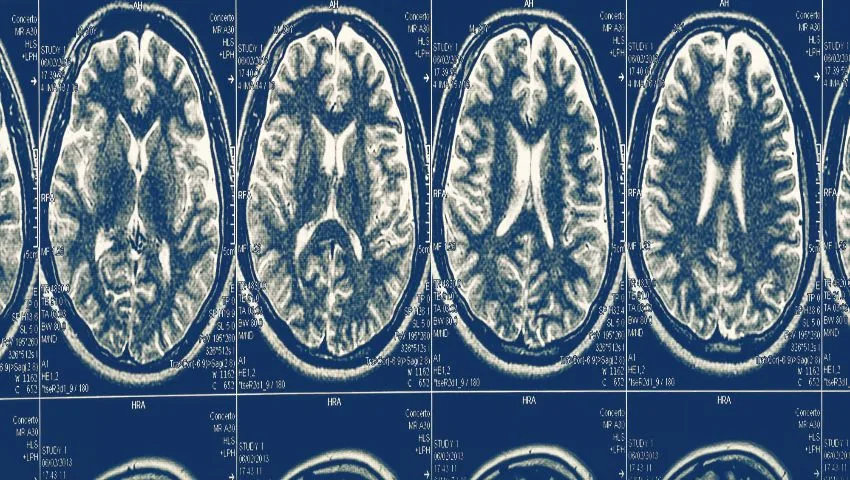
- MRI - to evaluate spinal cord compression, cord signal changes (myelopathy), Chiari malformation and syringomyelia.
- Additional tests - blood tests for inflammatory disease or infection, and assessment of swallowing and breathing in patients with brainstem symptoms.
Treatment of Atlanto-Axial Dislocation
The goals of treatment are to restore alignment, relieve pressure on the spinal cord and brainstem and achieve lasting stability so that further damage is prevented.
Non-surgical management
Selected patients - for example, children with acute rotatory subluxation, some stable fractures and asymptomatic mild instability - may be managed with medicines, a cervical collar, traction or a halo brace, with close follow-up imaging. However, established AAD with cord compression or significant instability generally requires surgery, as it does not heal on its own and carries a risk of sudden deterioration.
Surgical treatment
Modern surgery for AAD is based on the principle of direct joint manipulation and stable internal fixation. Commonly used techniques include:
C1 lateral mass - C2 pedicle/pars screw fixation
The most widely used technique. Screws are placed into C1 and C2 and connected with rods, allowing reduction and rigid stabilisation.
C1-C2 joint distraction and spacer
For irreducible AAD and basilar invagination, the joints are opened, distracted and packed with spacers or bone graft to realign the bones and pull the dens down.
Transarticular screws
Screws passing across the C1-C2 joints - effective where anatomy permits.
Occipito-cervical fusion
When C1 is assimilated with the skull or C1 fixation is not possible, the skull is fixed to the upper cervical spine.
Odontoid screw
For certain fresh odontoid fractures, a single screw placed from the front can fix the fracture while preserving rotation.
Transoral / endoscopic endonasal decompression
Rarely needed today; used when compression from the front cannot be relieved by posterior realignment.
The Surgical Procedure - Step by Step
- Detailed planning with 3D CT and CT angiography to measure screw trajectories and identify the vertebral artery course.
- Anaesthesia with careful airway management - neck movement is minimised; fibreoptic intubation may be used.
- Neuromonitoring - baseline motor and sensory signals are recorded and monitored throughout.
- Prone positioning with the head in a fixation frame; alignment is checked with fluoroscopy.
- Exposure of C1 and C2 through a midline incision at the back of the neck, protecting the venous plexus and the C2 nerve roots.
- Joint preparation and reduction - the C1-C2 joints are opened, cartilage removed and the bones realigned.
- Screw placement under fluoroscopy or navigation, followed by rod connection to lock the reduction.
- Bone grafting to achieve permanent fusion.
- Closure in layers, and application of a soft or hard collar as advised.
Risks and Safety Measures
CVJ surgery is technically demanding. Potential risks include injury to the vertebral artery, spinal cord or nerves, cerebrospinal fluid leak, infection, failure of fusion, hardware problems, difficulty swallowing and restriction of neck rotation after C1-C2 fusion (since this joint provides much of the head's rotation). Other neck segments usually compensate for much of this loss over time. These risks are minimised through meticulous planning, CT angiography, navigation, neuromonitoring and experience - factors that make the choice of surgeon particularly important for this condition.
Recovery After AAD Surgery
General recovery pathway
- First 24-48 hours: monitoring in a high-dependency or ICU setting; sitting up and early mobilisation with support.
- Hospital stay: usually several days, depending on neurological status and the complexity of surgery.
- Collar: a cervical collar may be advised for a period of weeks.
- Physiotherapy: gait training, balance work and hand function exercises.
- Follow-up imaging: X-rays and CT to confirm alignment and fusion over the following months.
Neurological recovery depends on how severe and how long-standing the cord compression was before surgery. Many patients notice improvement in walking, hand function and neck pain over weeks to months. Even when full recovery is not possible, stabilisation prevents further deterioration and protects against sudden injury.
AAD in Children
Children with congenital CVJ anomalies, Down syndrome or skeletal dysplasias need special attention. Screening X-rays may be recommended before participation in certain sports. Surgery in children uses techniques adapted to smaller, growing bones, and families are closely involved in decisions and rehabilitation. Early treatment in symptomatic children can prevent lifelong disability.
AAD in the Elderly - Odontoid Fractures
Older adults can fracture the dens after a simple fall. These fractures are sometimes missed because the neck pain seems mild. Treatment depends on the fracture type, bone quality and general health - options range from collar immobilisation to odontoid screw fixation or posterior C1-C2 fusion. Timely diagnosis in any elderly person with neck pain after a fall is important.
Why Choose Dr. (Prof.) Sumiet Snha for CVJ and AAD Surgery?
- Trained and taught at AIIMS, New Delhi - one of the world's highest-volume centres for craniovertebral junction anomalies.
- MBBS, MS, DNB, MCh (AIIMS), FACS, with over 25 years of neurosurgical experience.
- Former Professor of Neurosurgery at AIIMS and founder faculty in-charge of its cadaver training facility, where surgeons practise complex approaches like CVJ fixation.
- Certified AO Spine International faculty and ATLS faculty with deep experience in spinal trauma.
- Use of CT angiography-based planning, navigation and neuromonitoring for maximum safety.
- Comprehensive care for children, adults and the elderly at Max Super Speciality Hospital, Dwarka.
Factors That Influence the Cost of AAD Surgery
- Type of AAD (reducible vs irreducible, with or without basilar invagination).
- Fixation technique, implants and use of navigation.
- Need for ICU care and length of hospital stay.
- Associated procedures such as Chiari decompression.
- Patient's general health and insurance coverage.
Associated Conditions: Chiari Malformation and Syringomyelia
AAD with basilar invagination is frequently accompanied by Chiari malformation, in which the lower part of the cerebellum (tonsils) descends into the upper spinal canal, and by syringomyelia, a fluid-filled cavity within the spinal cord. Patients may experience headaches that worsen with coughing or straining, loss of pain and temperature sensation in a "cape-like" distribution over the shoulders and arms, hand wasting and scoliosis. Research, much of it from Indian centres, has shown that in many such patients the underlying problem is instability at the C1-C2 joints, and that realigning and fixing these joints can relieve both the bony compression and the associated Chiari and syrinx. The choice between joint fixation, foramen magnum decompression or a combination is individualised based on imaging and symptoms.
Living With a Stabilised Neck
After successful C1-C2 fusion, most patients return to their daily routines, school or work. Rotation of the head is somewhat reduced, but people quickly learn to compensate by turning the shoulders slightly, and other neck joints gradually take up some of the movement. Driving is usually possible once the surgeon confirms healing and the patient can check blind spots safely - wider mirrors can help. Contact sports and activities with a risk of heavy falls may be restricted depending on the extent of fusion. Regular follow-up allows the surgeon to confirm solid fusion and monitor the adjacent segments. Good posture, neck-strengthening exercises taught by the physiotherapist and avoidance of prolonged forward bending of the head (for example, over mobile phones) help maintain comfort in the long term.
Home Care and Rehabilitation After Surgery
- Collar care: wear the collar exactly as advised; keep the skin under it clean and dry, and check for pressure marks, especially on the chin and back of the head.
- Wound care: keep the incision dry until the surgeon permits bathing; report redness, discharge or fever.
- Posture: sit upright with support; avoid looking down for long periods and bring screens to eye level.
- Walking: walk several times a day, using a walker or stick initially if balance is affected.
- Hand exercises: squeezing a soft ball, picking up small objects and practising buttoning improve dexterity.
- Swallowing: take small bites and sips in an upright position; mention any choking or voice change.
- Sleep: use a thin pillow that keeps the neck in a neutral position.
Questions Families Often Ask Before Surgery
Families understandably want to know whether surgery is truly necessary, what would happen without it, and what life will look like afterwards. In patients with documented instability and cord compression, the natural course is often gradual worsening punctuated by sudden setbacks after minor injuries, which is why stabilisation is usually recommended. Surgery aims first to protect and then to improve neurological function. Dr. Snha takes time to explain the scans, the planned screws and joint realignment, the expected hospital stay and the realistic recovery timeline, so that families can make a confident, informed decision.
How Imaging Measurements Guide Treatment
Several measurements on X-ray, CT and MRI help the surgeon judge how serious the instability is. The atlanto-dental interval (ADI) is the gap between the front arch of the atlas and the dens; in adults it is normally no more than about 3 mm, and an increase suggests ligament failure. The posterior atlanto-dental interval, or space available for the cord, reflects how much room is left for the spinal cord behind the dens - a narrowing of this space is closely related to the risk of neurological injury. Lines drawn from the hard palate to the back of the foramen magnum (Chamberlain's line) and related reference lines help detect basilar invagination. The clivo-axial angle indicates how sharply the brainstem is bent over the dens. On MRI, a bright signal within the spinal cord indicates cord injury or swelling (myelomalacia). Taken together with the patient's symptoms, these measurements determine whether observation, a collar or surgical stabilisation is the right course, and which surgical technique will best restore alignment.
Because CVJ anatomy varies so much between individuals, no two operations are exactly alike. Detailed three-dimensional planning allows the surgeon to choose screw entry points, lengths and angles in advance, anticipate the position of the vertebral arteries and decide whether joint distraction will be needed to achieve reduction. This preparation shortens operating time and adds an important margin of safety.
Red-Flag Symptoms That Need Urgent Attention
People who have been diagnosed with atlanto-axial instability, or who are waiting for surgery, should know which symptoms mean the spinal cord or brainstem may be under increasing pressure. Because the upper neck houses the pathways controlling breathing, limb movement and swallowing, a sudden change should never be ignored or treated only with painkillers. Families are encouraged to keep the patient's scan reports and contact numbers easily accessible so that emergency doctors can be informed about the diagnosis straight away.
Seek emergency care if you notice
New or rapidly worsening weakness in the arms or legs; sudden numbness or electric-shock sensations down the body on bending the neck; difficulty walking or frequent falls; new problems with bladder or bowel control; difficulty swallowing, change in voice or breathing difficulty; or episodes of dizziness, double vision or fainting with neck movement.
After any fall or accident, a person with known AAD should keep the neck still and inform the emergency team of the diagnosis before being moved or intubated. Minor-looking trauma can cause disproportionate harm when the upper neck is already unstable.
Seeking a Second Opinion on a CVJ Diagnosis
Craniovertebral junction problems are relatively uncommon, and the recommended treatment can differ depending on how the images are interpreted. It is entirely reasonable for patients to seek a second opinion before major surgery. A useful review needs more than the written report: the original CT images, MRI of the cervical spine and, ideally, flexion-extension X-rays or dynamic CT showing whether the joint is reducible. A CT angiogram, if already done, helps assess the course of the vertebral arteries.
Dr. Snha, as a former Professor of Neurosurgery at AIIMS New Delhi and AO Spine International faculty, regularly reviews complex CVJ cases referred from other cities and countries. Patients can send their images and reports on WhatsApp for a preliminary opinion, and a video consultation can be arranged. When neurological symptoms are progressing, however, the review should be done urgently so that treatment is not delayed.
Frequently Asked Questions
Is atlanto-axial dislocation dangerous?
It can be. Because the spinal cord at this level controls breathing and all four limbs, unstable AAD can cause progressive or sudden neurological deficit. Timely evaluation and treatment by an experienced neurosurgeon are important.
Can AAD be treated without surgery?
Some cases, such as acute rotatory subluxation in children or certain stable fractures, can be treated with a collar or traction. Established AAD with cord compression usually requires surgical fixation.
Will I be able to move my neck after C1-C2 fusion?
Nodding movement is preserved and other neck segments compensate partly for the reduced rotation. Most patients adapt well and carry out normal daily activities.
How long does recovery take after AAD surgery?
Hospital stay is usually several days. Many patients resume light activities within a few weeks, while bone fusion takes around three to six months. Neurological recovery can continue for many months.
Is AAD surgery safe in children?
Yes, with appropriate planning and techniques adapted to the growing spine. Early treatment in symptomatic children can prevent serious long-term disability.
What activities should I avoid if I have AAD?
Avoid neck manipulation, forceful massage, contact sports, diving and sudden jerky neck movements until the condition is treated and your surgeon advises otherwise.
Why is a CT angiogram needed before surgery?
The vertebral arteries often follow unusual paths in CVJ anomalies. CT angiography maps them precisely so that screws can be placed safely.
Protect Your Spinal Cord - Consult an Expert
If you have been diagnosed with atlanto-axial dislocation, basilar invagination or another craniovertebral junction anomaly - or have neck pain with weakness or imbalance - early expert evaluation is essential. Book a consultation with Dr. (Prof.) Sumiet Snha at Max Hospital, Dwarka, New Delhi. Related reading: spinal fractures, paediatric neurosurgery and cervical and lumbar stenosis.