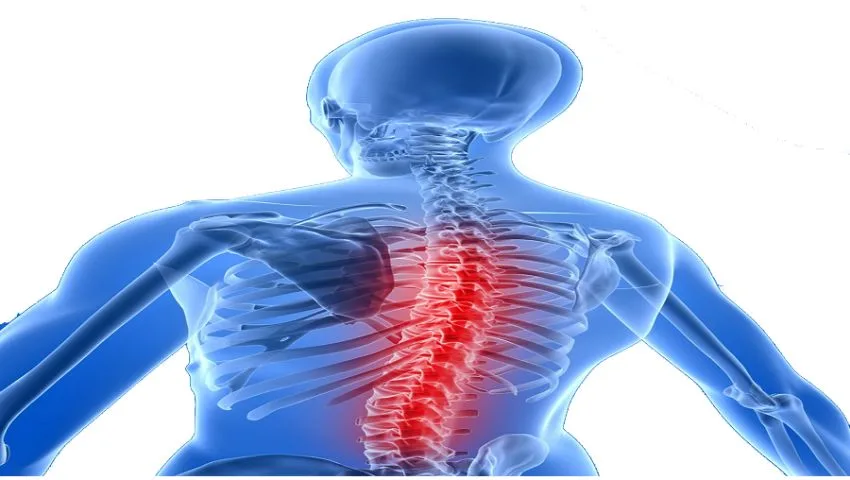
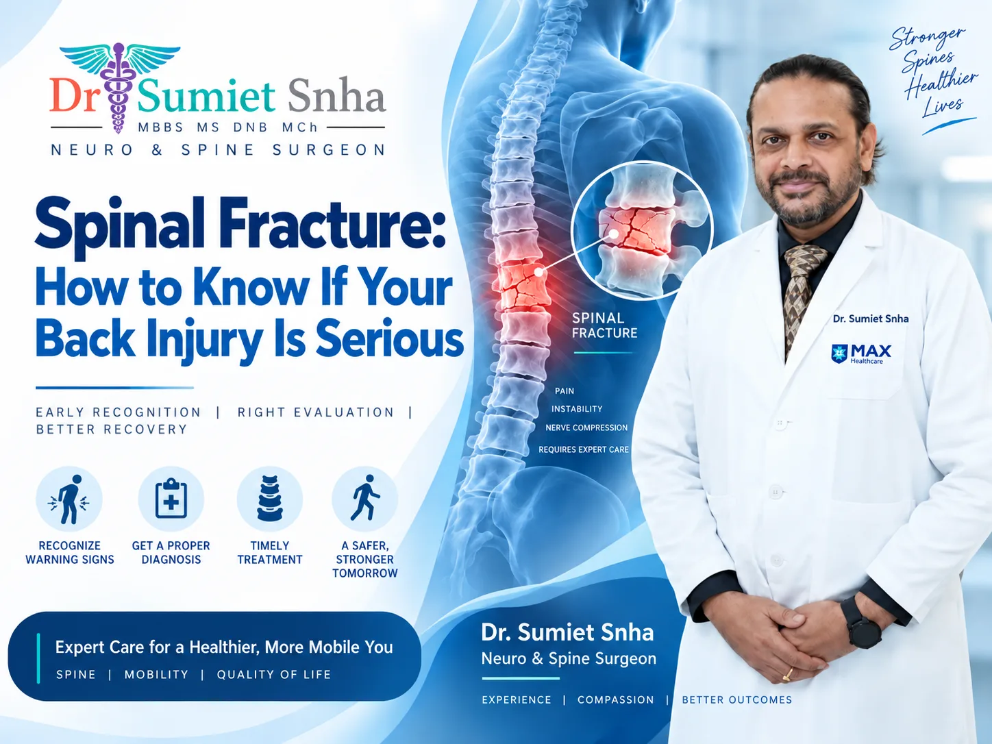
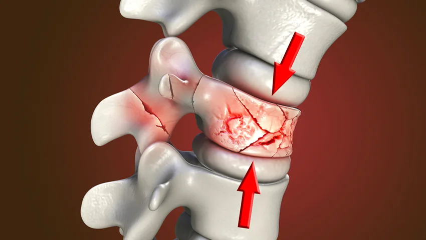
A fall on the stairs, a road traffic accident, a slip in the bathroom or, in someone with weak bones, even a cough or lifting a bucket - any of these can fracture a bone in the spine. Spinal fractures are different from fractures of the arm or leg. The spine not only supports the body's weight but also protects the spinal cord and nerve roots, so a fracture can affect stability, cause severe pain and, in some cases, injure the nerves. Some fractures heal well with rest and a brace; others need prompt surgery to prevent deformity or paralysis.
Dr. (Prof.) Sumiet Snha has special training and long experience in spinal injuries. A former Professor of Neurosurgery at AIIMS, New Delhi - including at the JPNA Apex Trauma Centre - he is a certified teaching faculty for AO Spine International and a certified ATLS (Advanced Trauma Life Support) faculty, and has served as Secretary and later President of the Neurotrauma Society of India. He was also a member of the core committee that formulated national guidelines for traumatic brain injury. At Max Hospital, Dwarka, New Delhi, he treats the full spectrum of spinal fractures - from osteoporotic compression fractures in the elderly to complex fracture-dislocations after high-energy trauma.
Understanding the Spine and How It Fractures
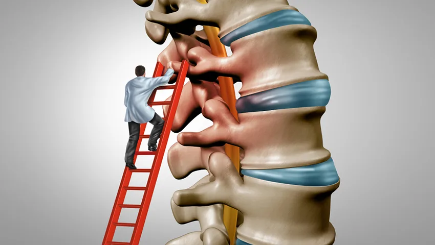
Your spine is made up of 33 vertebrae - 7 in the neck (cervical), 12 in the upper and mid-back (thoracic), 5 in the lower back (lumbar), and the fused sacrum and coccyx. Each vertebra has a solid body in front that carries weight and a bony arch behind that surrounds and protects the spinal canal. Ligaments, discs and muscles hold the column together, allowing movement while maintaining stability.
Surgeons often think of the spine as having three columns: the anterior column (front half of the vertebral body and disc), the middle column (back half of the body, next to the spinal canal) and the posterior column (the bony arch and ligaments at the back). Fractures that involve only the front column are usually stable, while those that break through two or three columns, or tear the posterior ligaments, are more likely to be unstable. Most fractures occur at the thoracolumbar junction (around T11-L2), where the relatively rigid chest spine meets the more mobile lower back and forces concentrate.
Types of Spinal Fractures
Compression fracture
The front of the vertebral body collapses into a wedge shape. Very common in osteoporosis; usually stable.
Burst fracture
The vertebral body shatters under a heavy axial load, and fragments may be pushed backwards into the spinal canal.
Flexion-distraction (Chance) fracture
The vertebra is pulled apart, typically in a car accident with a lap belt. Often unstable, with associated abdominal injuries.
Fracture-dislocation
The most severe type, in which vertebrae are displaced relative to each other. Often associated with spinal cord injury and needs surgery.
Transverse and spinous process fractures
Small fractures of the bony projections, usually stable and treated with pain relief and activity modification.
Pathological fracture
A fracture through bone weakened by a tumor, infection or metabolic disease, often after minimal trauma.
Modern classification systems, such as the AO Spine classification, grade fractures according to their morphology, neurological status and patient-specific factors. This helps surgeons decide objectively whether a fracture can be treated with a brace or needs surgery.
Causes of Spinal Fractures
High-energy trauma
Road traffic accidents, falls from height (for example, from roofs or construction sites), sports injuries and assault are the main causes in younger adults. These fractures may be associated with head, chest, abdominal and limb injuries, and patients need comprehensive trauma care.
Osteoporosis
Osteoporosis thins and weakens the bones, particularly in post-menopausal women and older men. In such people, a fracture can occur after a minor fall, a sudden twist, lifting a light object or even a bout of coughing. Many osteoporotic fractures go unnoticed or are dismissed as "ordinary back pain". Multiple compression fractures can gradually lead to loss of height and a stooped posture (kyphosis).
Tumors and infection
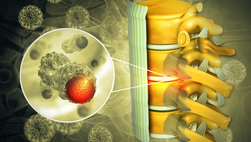
Cancer that has spread to the spine, multiple myeloma and spinal infections such as tuberculosis can destroy bone and cause fractures with little or no injury. A fracture without a clear cause always needs investigation for an underlying disease.
Other risk factors
- Long-term steroid use.
- Rheumatoid arthritis and ankylosing spondylitis - in the latter, the fused, rigid spine can break like a long bone after minor trauma.
- Vitamin D deficiency, low body weight, smoking and excess alcohol.
- Chronic kidney or liver disease and certain hormonal disorders.
Symptoms of a Spinal Fracture
- Sudden, severe back or neck pain after an injury, worse on movement, standing or walking.
- Pain that eases on lying down, typical of compression fractures.
- Localised tenderness over the spine.
- Muscle spasm and difficulty turning in bed or getting up.
- Loss of height or a new forward stoop (with multiple fractures).
- Numbness, tingling or weakness in the arms or legs if the nerves or spinal cord are affected.
- Bladder or bowel dysfunction in severe injuries.
After an accident - first aid matters
If someone has had a significant fall or accident and complains of neck or back pain, numbness or weakness, do not move them unnecessarily. Keep the head, neck and body in line, call for emergency help and allow trained personnel to immobilise the spine before transfer. Incorrect handling can turn a stable injury into a spinal cord injury.
How Spinal Fractures Are Diagnosed
- Trauma assessment - in accident victims, a systematic evaluation following ATLS principles to identify all injuries, protecting the spine throughout.
- Neurological examination - assessment of strength, sensation and reflexes, recorded using a standard scale to document the level and severity of any spinal cord injury.
- X-rays - the initial test to identify fractures and alignment; standing X-rays help assess stability in selected cases.
- CT scan - the best test to define bony fractures, fragments in the canal and the pattern of injury.
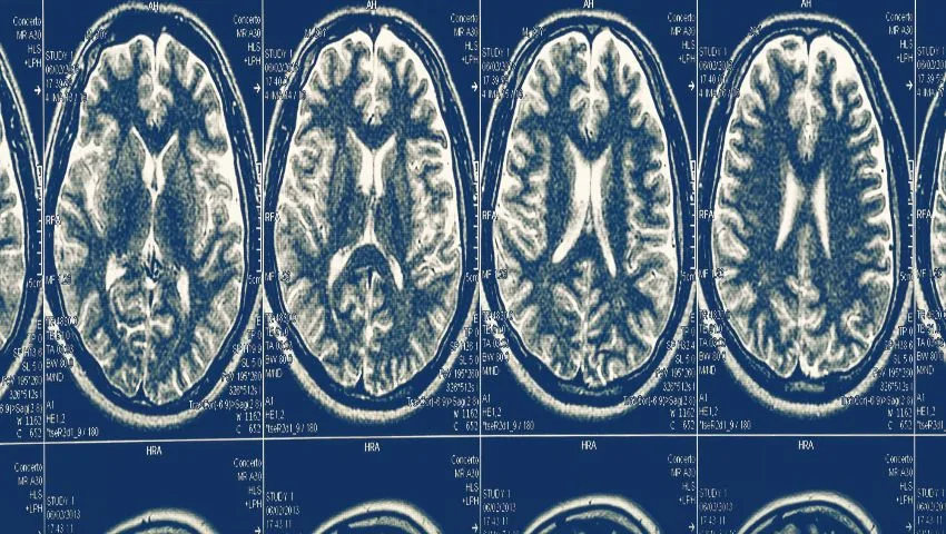
- MRI scan - to assess the spinal cord, nerve roots, discs and ligaments, and to judge whether a compression fracture is new or old (fresh fractures show bone marrow swelling).
- Bone density scan (DEXA) - to diagnose osteoporosis after a low-energy fracture.
- Blood tests and biopsy - when a tumor or infection is suspected.
Treatment Options for Spinal Fractures
The aims of treatment are to relieve pain, protect and restore nerve function, maintain or restore alignment and stability, and return the patient to activity as quickly and safely as possible.
Non-surgical treatment
Most stable fractures, including many compression fractures and some burst fractures without nerve involvement, can be treated without surgery:
Pain management
Appropriate painkillers to allow early mobilisation, with care in elderly patients.
Bracing
A custom or off-the-shelf brace (TLSO or cervical collar) for a period of weeks to support the spine while it heals.
Early mobilisation
Walking with support as soon as pain allows, to prevent complications of bed rest such as clots, chest infection and muscle wasting.
Osteoporosis treatment
Calcium, vitamin D and bone-strengthening medicines to reduce the risk of further fractures.
Regular follow-up X-rays check that the fracture is not collapsing further or tilting the spine.
Vertebroplasty and kyphoplasty
For painful osteoporotic or tumor-related compression fractures that do not settle with conservative care, vertebral augmentation can provide rapid pain relief. In vertebroplasty, medical bone cement is injected into the fractured vertebra through a needle under X-ray guidance. In kyphoplasty, a small balloon is first inflated inside the vertebra to create a cavity and partially restore height, and the cavity is then filled with cement. Both are done through tiny puncture wounds, often under local anaesthesia with sedation, and many patients are able to walk within hours with noticeably less pain.
Minimally invasive percutaneous fixation
For unstable fractures without significant nerve compression, screws can be inserted into the vertebrae above and below the fracture through small stab incisions and connected with rods passed under the skin. This stabilises the fracture with minimal muscle damage and blood loss, allows early mobilisation and avoids prolonged bed rest or bracing. In some cases, the screws can be removed after the fracture has healed.
Open decompression and fixation
When bone fragments or a displaced vertebra are compressing the spinal cord or nerve roots, surgery is performed to remove the pressure (decompression), realign the spine and stabilise it with screws and rods. Depending on the injury, this may be done from the back, the front or both. In severe burst fractures, the crushed vertebral body may be replaced with a cage (corpectomy).
Cervical spine fractures
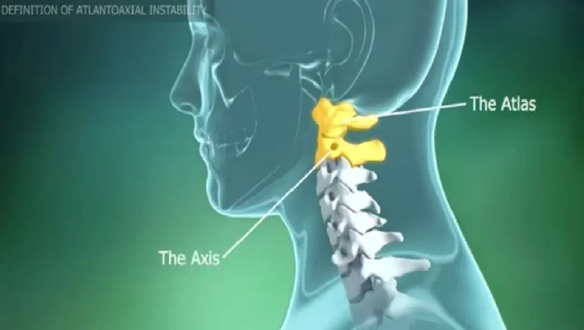
Neck fractures are treated according to their level and type - with a collar, a halo device or surgical fixation from the front (anterior cervical discectomy/corpectomy and fusion) or the back (lateral mass or pedicle screw fixation). Injuries of the upper cervical spine (C1-C2) are discussed in detail on our atlanto-axial dislocation page.
When Is Surgery Needed?
Surgery is generally recommended when:
- The spinal cord or nerve roots are compressed and there is neurological deficit, especially if it is worsening.
- The fracture is unstable - for example, fracture-dislocations, flexion-distraction injuries and burst fractures with disruption of the posterior ligaments.
- There is significant deformity or progressive collapse despite bracing.
- Pain remains severe and disabling despite adequate conservative treatment.
- The fracture is caused by a tumor or infection requiring tissue diagnosis and stabilisation.
- Early mobilisation is important to avoid complications, as in polytrauma patients or the elderly.
Spinal Cord Injury - Acting Fast
When a fracture injures the spinal cord, time is critical. Early decompression and stabilisation, ideally within the first hours after injury when the patient's overall condition allows, may improve the chances of neurological recovery. Maintaining good blood pressure and oxygen levels to protect the injured cord is also important. The extent of recovery depends mainly on the severity of the initial injury: patients with incomplete injuries (some preserved sensation or movement below the level of injury) have a significantly better outlook than those with complete injuries. Comprehensive rehabilitation - physiotherapy, occupational therapy, bladder and bowel management, skin care and psychological support - is an essential part of treatment. In selected patients with cervical spinal cord injury, later reconstructive procedures such as nerve transfers may help restore some hand and arm function.
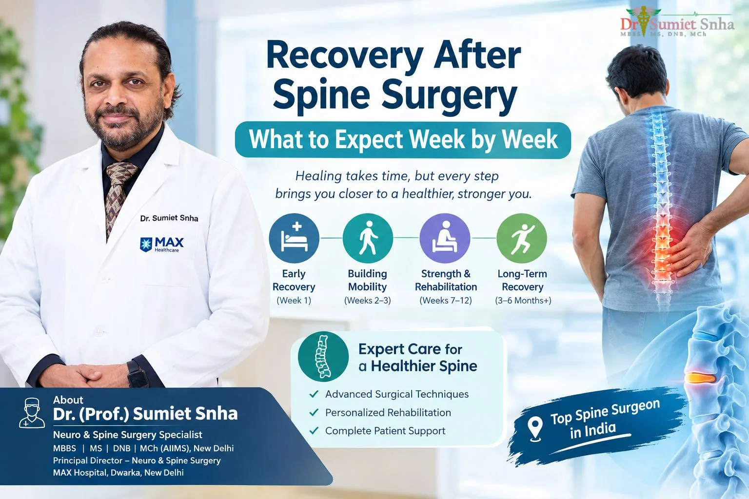
Recovery After Spinal Fracture Treatment
General timeline
- Kyphoplasty / vertebroplasty: walking within hours; most patients go home within a day and resume light activities within days.
- Percutaneous fixation: walking within one to two days; discharge in a few days; return to desk work in a few weeks.
- Open decompression and fusion: hospital stay of several days; structured rehabilitation over weeks to months.
- Bracing (non-surgical): usually 8-12 weeks with periodic X-rays, followed by strengthening exercises.
- Bone healing: typically takes around three months; fusion may take longer.
Physiotherapy focuses on posture, core and back extensor strength, balance and safe movement patterns. Elderly patients benefit from fall-prevention measures at home, such as good lighting, grab bars in the bathroom, non-slip mats and appropriate footwear.
Osteoporotic Fractures - Treating the Bone, Not Just the Break
A spinal compression fracture after a minor fall is often the first sign of osteoporosis. One fracture significantly increases the risk of another, a phenomenon sometimes called the "fracture cascade". Therefore, treating the fracture is only half the job. Every patient with a low-energy spinal fracture should have a bone density assessment and appropriate treatment, which may include calcium and vitamin D supplements, bone-building or bone-protecting medicines, weight-bearing exercise, stopping smoking, limiting alcohol and measures to prevent falls. Coordinating with an endocrinologist or physician ensures long-term bone health.
Possible Complications
Without appropriate treatment, spinal fractures may lead to chronic pain, progressive deformity (kyphosis), loss of height, reduced lung capacity with multiple thoracic fractures, and neurological deterioration. Surgical treatment carries its own risks - infection, bleeding, nerve injury, implant problems, cement leakage in vertebral augmentation and failure of fusion - which are minimised through careful technique and are discussed openly before any procedure.
Why Choose Dr. (Prof.) Sumiet Snha for Spinal Fracture Care?
- Specialist in spinal trauma - former Professor of Neurosurgery at AIIMS, New Delhi, with extensive work at the JPNA Apex Trauma Centre.
- President, Neurotrauma Society of India, and earlier its Secretary.
- Certified AO Spine International faculty and ATLS faculty - teaching surgeons and doctors the latest standards in spine and trauma care.
- Member of the core committee that formulated national guidelines for traumatic brain injury.
- Expertise across the full range of treatment - bracing, kyphoplasty, minimally invasive fixation and complex reconstruction.
- Round-the-clock emergency and trauma infrastructure at Max Super Speciality Hospital, Dwarka, New Delhi.
What Influences the Cost of Spinal Fracture Treatment?
- Type and level of fracture and whether nerves are involved.
- Choice of treatment - brace, vertebroplasty/kyphoplasty, percutaneous fixation or open surgery.
- Number of levels fixed and implants used.
- ICU requirements and hospital stay, especially after polytrauma.
- Rehabilitation needs.
- Insurance coverage, including accident policies.
Preventing Spinal Fractures
- Always wear seat belts and helmets; follow safe driving practices.
- Use safety harnesses when working at height.
- Keep bones strong with a calcium-rich diet, adequate vitamin D and regular weight-bearing exercise.
- Get a bone density test if you are a post-menopausal woman, a man over 70, or have other risk factors.
- Make your home fall-safe: remove loose rugs, install handrails and bathroom grab bars, and ensure good lighting at night.
- Review medicines that cause dizziness or drowsiness with your doctor.
- Avoid diving into shallow water.
Living With a Healed Spinal Fracture
Once a fracture has healed, most people return to their normal activities. Some may have mild residual back ache, particularly after prolonged standing, which usually responds well to regular exercise and posture awareness. Those who had multiple osteoporotic fractures benefit from ongoing bone health treatment and exercise programmes that emphasise back extensor strength and balance. Patients who had spinal cord injury continue rehabilitation and periodic reviews to maximise independence and prevent complications. At every stage, clear communication between patient, family and the treating team leads to the best outcomes.
Special Situations in Spinal Fracture Care
Fractures in ankylosing spondylitis and DISH
In ankylosing spondylitis and diffuse idiopathic skeletal hyperostosis (DISH), the spine gradually fuses into a rigid column. A rigid spine behaves like a long bone: it cannot absorb forces by bending, so even a minor fall can fracture it straight through, often across the disc space. These fractures are highly unstable, are easily missed on plain X-rays and carry a significant risk of spinal cord injury. Any person with ankylosing spondylitis who develops new neck or back pain after even a trivial injury should have a CT scan, and many such fractures are best treated with surgical stabilisation spanning several levels above and below the break.
Sacral insufficiency fractures
Elderly people with osteoporosis, and those who have had radiation to the pelvis, can develop stress fractures of the sacrum - the triangular bone at the base of the spine. The pain is felt low in the back, buttocks or groin and is often mistaken for a slip disc or hip arthritis. MRI or CT confirms the diagnosis. Most heal with pain relief, protected walking and osteoporosis treatment; persistent cases may benefit from sacroplasty (cement injection) or fixation.
Spinal injuries in children and adolescents
Children's spines are more flexible, and injury patterns differ from adults. Young children are more prone to upper cervical injuries because of their relatively large heads, while adolescents injured in sports or falls tend to have adult-type fractures. Occasionally, the spinal cord can be injured without an obvious fracture on X-ray, which is why MRI is important in any child with neurological symptoms after trauma. Treatment respects the growing spine, and most children recover very well from stable injuries.
Polytrauma patients
After major accidents, spinal fractures often coexist with head, chest, abdominal and limb injuries. Treatment is coordinated by a trauma team so that life-threatening injuries are addressed first while the spine is protected. Early stabilisation of spinal fractures, when the patient is fit, helps nursing care, reduces lung complications and allows earlier rehabilitation.
Returning to Work, Driving and Sport
The timing of return to normal life depends on the fracture type, the treatment and the demands of the activity. As a broad guide, people with desk-based jobs can often return within two to six weeks after kyphoplasty or minimally invasive fixation, whereas physically demanding work may require three months or more. Driving can usually be resumed when you no longer need strong painkillers, can sit comfortably for the length of the journey, and can turn and brake safely - typically a few weeks after minimally invasive surgery. Sports are reintroduced gradually after healing is confirmed, starting with low-impact activities such as walking, swimming and cycling, and progressing to contact or high-impact sports only on the surgeon's advice. A physiotherapist can design a return-to-activity programme that rebuilds strength and confidence.
Psychological Recovery
A spinal injury can be emotionally as well as physically challenging. Pain, time away from work, dependence on others and, after serious accidents, memories of the event can affect mood and sleep. Feeling anxious or low is common and is nothing to be ashamed of. Talking openly with the treating team, involving family, connecting with other patients and, where needed, seeking help from a counsellor or psychologist all support a fuller recovery. For patients with spinal cord injury, peer support and vocational rehabilitation play a key role in rebuilding independence and purpose.
Glossary
- Kyphosis: a forward curvature or hunching of the spine.
- Retropulsion: backward displacement of bone fragments into the spinal canal.
- Corpectomy: removal of a vertebral body, usually replaced with a cage.
- Pedicle screw: a screw placed through the pedicle (bony bridge) into the vertebral body.
- TLSO: thoraco-lumbo-sacral orthosis - a brace supporting the mid and lower back.
- Incomplete spinal cord injury: an injury in which some movement or sensation is preserved below the level of injury.
Nutrition for Faster Bone Healing
Bones need building material to heal. A balanced diet with adequate protein - from dal, pulses, milk, curd, paneer, eggs, fish or lean meat - supports the formation of new bone and helps maintain muscle strength during recovery. Calcium-rich foods such as milk products, ragi, sesame seeds and green leafy vegetables, together with sufficient vitamin D from sunlight or supplements, are essential for mineralising the healing bone. Vitamin D deficiency is extremely common in India, including in people who spend little time outdoors, and correcting it is a simple but important step. Smoking should be stopped completely, as nicotine reduces blood flow to bone and significantly delays fracture healing and spinal fusion. Alcohol should be limited. Patients with diabetes should keep their blood sugar well controlled, since high sugar levels slow healing and raise the risk of infection. Your doctor may recommend specific supplements based on blood tests.
Staying as active as the fracture allows is also part of healing. Gentle walking improves circulation, prevents stiffness and stimulates bone repair. Follow your surgeon's and physiotherapist's guidance about how much activity is safe at each stage, and do not hesitate to ask if you are unsure.
Managing Pain While a Spinal Fracture Heals
Good pain control is not just about comfort - it allows patients to sit, walk, breathe deeply and take part in physiotherapy, all of which reduce the risk of chest infection, blood clots and muscle loss. The aim is to use the smallest effective dose of medicines for the shortest necessary time, adjusting the plan as the fracture heals. Pain that suddenly increases, rather than gradually settling, should be reported, as it may signal further collapse of the vertebra or another problem that needs review.
Common approaches
- Simple analgesics: paracetamol taken regularly is often the foundation of treatment.
- Anti-inflammatory medicines: helpful for some patients but used cautiously in the elderly and in those with kidney, stomach or heart problems.
- Stronger painkillers: sometimes needed for a short period early on, with a plan to reduce them; constipation and drowsiness need attention, especially in older people.
- Nerve pain medicines: used when a fracture irritates a nerve root, producing burning or shooting pain.
- Bracing and positioning: a well-fitted brace and advice on getting in and out of bed reduce pain on movement.
- Cold or warm packs and gentle mobilisation: guided by the physiotherapist.
When pain does not settle
Most stable fractures become noticeably less painful over four to six weeks. Pain that remains severe beyond this, or that keeps a person bed-bound, may indicate that the fracture is not healing, is collapsing further, or that the vertebra contains fluid or a gap on MRI. In such cases, vertebral augmentation or fixation may be reconsidered. Dr. Snha reviews persistent pain with repeat imaging rather than simply increasing medicines, because treating the mechanical cause is usually more effective and safer in the long run, particularly for older patients.
Frequently Asked Questions
Can a spinal fracture heal on its own?
Many stable fractures, such as most compression fractures, heal with pain relief, a brace and gradual activity over about three months. Unstable fractures or those affecting the nerves usually need surgery.
How do I know if my back pain is from a fracture?
Sudden, severe back pain after a fall or injury - or even after a minor strain in someone with osteoporosis - should be checked with an X-ray or MRI. Pain that is worse on standing and relieved by lying down is typical of compression fractures.
Is kyphoplasty safe for elderly patients?
Kyphoplasty and vertebroplasty are minimally invasive and are commonly performed in elderly patients with painful osteoporotic fractures. Suitability is decided after clinical assessment and MRI.
Will I be able to walk after spinal fracture surgery?
Most patients without spinal cord injury walk within one or two days after minimally invasive fixation. In spinal cord injury, recovery depends on the severity of the initial injury.
How long should I wear a brace?
Usually 8 to 12 weeks for thoracolumbar fractures treated conservatively, depending on healing seen on follow-up X-rays.
Do screws need to be removed later?
Not usually. In selected young patients with percutaneous fixation, screws may be removed after the fracture heals to restore segment mobility.
How can I prevent another osteoporotic fracture?
Treat the underlying osteoporosis with medicines, vitamin D and calcium, keep active with weight-bearing exercise, stop smoking and make your home fall-safe.
Get Expert Spinal Fracture Care
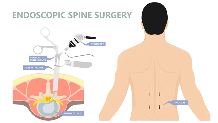
Whether it is a painful compression fracture after a fall or a serious injury after an accident, timely assessment by an experienced spine surgeon makes a difference. Book an appointment with Dr. (Prof.) Sumiet Snha at Max Hospital, Dwarka, New Delhi. You may also find these pages helpful: endoscopic spine surgery, spinal tuberculosis and spinal tumors.