A diagnosis of a brain tumor is one of the most frightening news a person can receive. Questions come rushing in: Is it cancer? Can it be removed? Will I be the same person after surgery? The reassuring reality is that brain tumors are a very diverse group of conditions, and many of them - including a large proportion of meningiomas, pituitary adenomas, schwannomas and other benign tumors - can be removed with excellent results. Even for malignant tumors, modern surgery combined with radiation and medical therapy has significantly improved outcomes and quality of life.
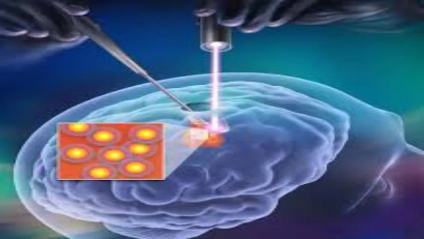
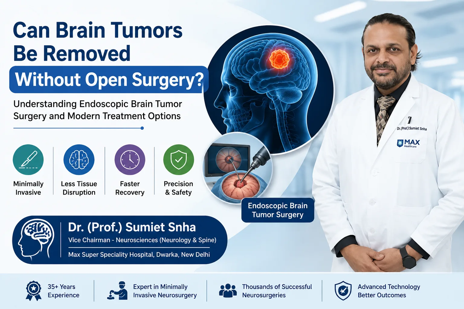
Brain tumor surgery has changed dramatically over the past two decades. Large skull openings and extensive brain exposure have increasingly given way to endoscopic and keyhole (minimally invasive) approaches, guided by neuro-navigation and advanced imaging. These techniques allow surgeons to reach tumors through small openings - sometimes entirely through the nose - with less disturbance to healthy brain, less pain and faster recovery. Dr. (Prof.) Sumiet Snha, former Professor of Neurosurgery at AIIMS, New Delhi, and Principal Director - Neurosurgery & Spine Surgery at Max Hospital, Dwarka, performs endoscopic and keyhole removal of brain tumors, including pituitary and skull base tumors.
What Is a Brain Tumor?
A brain tumor is an abnormal growth of cells within the skull - in the brain tissue itself, its coverings (meninges), the nerves leaving the brain, the pituitary gland or the skull base. Tumors are broadly classified as:
Primary tumors
Arise within the brain or its coverings. Examples: gliomas, meningiomas, pituitary adenomas, schwannomas, craniopharyngiomas.
Secondary (metastatic) tumors
Spread to the brain from cancers elsewhere - commonly lung, breast, kidney, melanoma and colon.
Benign tumors
Non-cancerous, usually slow-growing and well-defined; can still cause serious problems by pressing on the brain.
Malignant tumors
Cancerous, tend to grow faster and infiltrate surrounding brain; graded from 1 to 4 by the WHO.
Because the skull is a closed box, even a benign tumor can raise pressure inside the head or compress vital structures. The type, location and size of the tumor determine symptoms and treatment.
Common Types of Brain Tumors
Meningioma
The most common primary brain tumor in adults, arising from the meninges. Most are benign and slow-growing. They can occur over the surface of the brain, along the midline, or at the skull base, where they may involve nerves and blood vessels. Complete removal often results in cure.
Glioma
Tumors arising from the glial (supporting) cells of the brain, ranging from low-grade gliomas, which grow slowly, to glioblastoma, the most aggressive form. Treatment involves maximal safe surgical removal followed by radiation and/or chemotherapy based on grade and molecular markers.
Pituitary adenoma
Usually benign tumors of the pituitary gland at the base of the brain. They may produce excess hormones (causing conditions such as acromegaly or Cushing's disease) or press on the optic nerves, causing vision loss. Most are ideally removed through the nose using the endoscope.
Vestibular schwannoma (acoustic neuroma)
Benign tumors of the balance nerve, causing hearing loss, tinnitus and imbalance. Detailed information is on our acoustic neuroma page.
Craniopharyngioma
Benign but challenging tumors near the pituitary and hypothalamus, seen in children and adults, often affecting vision and hormones.
Colloid cyst and intraventricular tumors
Tumors within the fluid spaces (ventricles) of the brain that can block fluid flow and cause hydrocephalus - often ideally treated endoscopically.
Brain metastases
The most common brain tumors overall in adults. Surgery, stereotactic radiosurgery or both are used depending on number, size and the patient's overall condition.
Symptoms of Brain Tumors
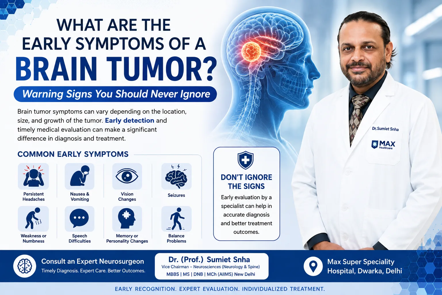
Symptoms depend on the tumor's location, size and speed of growth. Common symptoms include:
- Headaches - new, persistent or progressively worsening, often worse in the morning or on lying down, and sometimes accompanied by vomiting.
- Seizures (fits) - a first seizure in an adult should always be investigated.
- Weakness or numbness on one side of the body.
- Vision problems - blurred or double vision, loss of side vision.
- Speech difficulties - trouble finding words or understanding speech.
- Changes in personality, behaviour, memory or concentration.
- Balance problems, unsteadiness or clumsiness.
- Hearing loss or ringing in one ear.
- Hormonal changes - irregular periods, milk discharge, infertility, enlarging hands and feet, weight gain.
- Drowsiness or confusion in advanced cases.
When to seek urgent care
A sudden severe headache, a first seizure, sudden weakness, loss of vision, persistent vomiting with headache, or increasing drowsiness needs immediate medical attention. Most headaches are not due to tumors - but "red flag" headaches should always be evaluated. Read more in our blog on headaches that need a brain scan.
How Brain Tumors Are Diagnosed
- Neurological examination - assessment of strength, sensation, reflexes, coordination, vision, hearing and mental function.
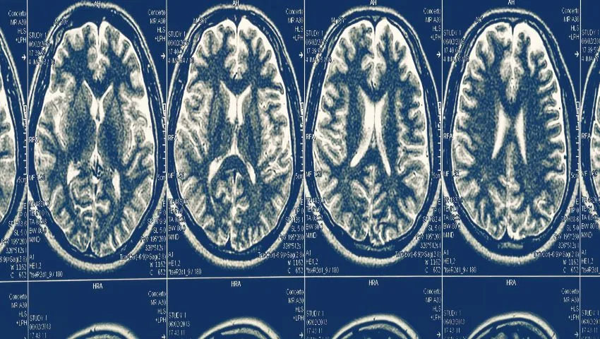
- MRI brain with contrast - the most important test, showing the tumor's size, location, relation to critical structures and characteristics that suggest its type.
- Advanced MRI - functional MRI (to map areas controlling movement and speech), diffusion tensor imaging or tractography (to map white matter pathways), perfusion and spectroscopy (to assess tumor grade).
- CT scan - for bone involvement, calcification and skull base anatomy.
- Hormone tests and eye examination - for pituitary and sellar tumors, including visual field testing.
- Whole-body evaluation - PET-CT when metastatic disease is suspected.
- Histopathology and molecular testing - analysis of the removed tissue confirms the diagnosis and guides further treatment.
Treatment Options
Treatment is individualised and often multidisciplinary:
Observation
Small, incidentally found benign tumors without symptoms may be monitored with periodic MRI.
Surgery
The mainstay for most symptomatic tumors - to remove the tumor, relieve pressure and obtain tissue for diagnosis.
Radiation / radiosurgery
Conventional radiotherapy or focused stereotactic radiosurgery for residual, recurrent or selected tumors.
Medical therapy
Chemotherapy, targeted therapy, and hormone-controlling medicines (e.g. for prolactinomas).
What Is Endoscopic Brain Tumor Surgery?
An endoscope is a slim tube with a light source and a high-definition camera at its tip. In neurosurgery, it provides a close-up, panoramic view of deep structures through a very small opening, allowing the surgeon to "look around corners" in a way the operating microscope cannot. Endoscopic brain tumor surgery comes in several forms:
Endoscopic endonasal (transsphenoidal) surgery
For pituitary adenomas and many skull base tumors, the surgeon passes the endoscope and instruments through the nostril into the sphenoid sinus, reaching the tumor from below - with no skin incision and no disturbance of the brain. Extended endonasal approaches can reach tumors of the anterior skull base, clivus and even some tumors above the pituitary. Patients typically recover quickly, with minimal pain and a short hospital stay.
Endoscopic intraventricular surgery
Tumors and cysts within the ventricles - such as colloid cysts - can be removed or biopsied through a small burr hole using a neuroendoscope. The same approach allows treatment of associated hydrocephalus through an endoscopic third ventriculostomy (see our hydrocephalus page).
Endoscope-assisted keyhole craniotomy
For tumors of the skull base and deep regions, the surgeon uses small, precisely placed openings - such as the supraorbital "eyebrow" approach or a small retrosigmoid opening behind the ear - combined with the endoscope and microscope. Planning with neuro-navigation ensures the opening is exactly where it needs to be.
Tubular retractor surgery
For deep-seated tumors within the brain, a narrow tubular port can be gently guided along natural pathways to the tumor, minimising disruption of surrounding brain.
Keyhole Surgery Principles
Keyhole neurosurgery is not simply about small incisions; it is about planning the smartest route to the tumor. By studying the MRI, the surgeon chooses an entry point that gives the best angle of view with the least brain retraction, often using natural corridors such as the nose, the space beneath the frontal lobe, or the angle between the cerebellum and brainstem. Endoscopes compensate for the smaller opening by bringing the light and camera close to the target. The result can be a comparable extent of tumor removal with less tissue trauma, when the approach is appropriate for the tumor. Not every tumor is suited to keyhole surgery - large, complex or highly vascular tumors may need a conventional approach - and experienced surgeons choose based on safety first.
Technology That Makes Brain Surgery Safer
- Neuro-navigation - a GPS-like system that shows the exact position of instruments on the patient's MRI in real time.
- High-definition and 3D endoscopes and operating microscopes for precise visualisation.
- Intra-operative neuromonitoring - monitoring of motor pathways and cranial nerves during surgery.
- Awake craniotomy with brain mapping - for tumors near speech or movement areas, the patient may be kept awake during part of the operation so that these functions can be protected.
- Fluorescence-guided surgery - special dyes can make certain tumors glow under specific light, helping distinguish tumor from brain.
- Ultrasonic aspirators and micro-instruments for gentle tumor removal.
- Intra-operative ultrasound or imaging to check the extent of removal.
The Brain Tumor Surgery Journey - Step by Step
- Consultation and planning - review of MRI and other tests; discussion of goals, options, risks and expected recovery with the patient and family.
- Pre-operative preparation - blood tests, anaesthesia assessment, hormone evaluation for pituitary tumors, and medicines such as steroids or anti-seizure drugs if needed.
- Surgery day - general anaesthesia (or awake surgery when indicated); navigation registration; the chosen endoscopic or keyhole approach; tumor removal; closure with careful reconstruction, especially of the skull base in endonasal surgery.
- Early recovery - monitoring in the neuro-ICU or high-dependency unit for the first night; a post-operative scan to assess removal.
- Ward care and mobilisation - most patients sit up and walk within a day or two; hospital stay is often a few days after minimally invasive procedures.
- Pathology and further plan - the tissue diagnosis determines whether additional treatment such as radiation is needed.
- Follow-up - clinical reviews and periodic MRI scans.
Benefits of Endoscopic and Keyhole Brain Tumor Surgery
- Smaller incisions and skull openings; in endonasal surgery, no visible scar.
- Less handling and retraction of healthy brain.
- Reduced post-operative pain and swelling.
- Shorter hospital stay and faster return to daily activities.
- Excellent visualisation of deep structures and tumor margins.
- Better cosmetic results - particularly important for young patients.
Risks of Brain Tumor Surgery
All brain surgery carries risks, which depend on the tumor's size, location and nature. Possible complications include bleeding, infection, seizures, cerebrospinal fluid leak (especially after endonasal surgery), new or worsened neurological deficits (weakness, speech, vision, hearing or balance problems), hormonal disturbances after pituitary surgery, and the anaesthetic risks of any major operation. Many deficits are temporary and improve over weeks. Your surgeon will explain the risks specific to your tumor, balanced against the risks of not treating it, so that you can make an informed decision.
Recovery After Brain Tumor Surgery
General recovery guide
- Hospital stay: often 2-5 days after endoscopic or keyhole surgery; longer after complex procedures.
- Endonasal surgery: avoid blowing the nose, straining and heavy lifting for a few weeks; nasal saline rinses help healing; some nasal congestion is normal.
- Craniotomy: keep the wound dry until advised; stitches or staples removed in about 10-14 days.
- Return to work: many patients return to desk work in 3-6 weeks, depending on the surgery and any further treatment.
- Driving: depends on the tumor type and whether seizures occurred - follow your doctor's advice.
- Rehabilitation: physiotherapy, occupational or speech therapy when needed.
Fatigue is common for several weeks after brain surgery. Gradually increasing activity, good sleep, balanced nutrition and staying hydrated help recovery. Report fever, clear fluid dripping from the nose, worsening headache, new weakness or seizures promptly.
Pituitary Tumors - A Closer Look
Pituitary adenomas deserve special mention because they are common and highly treatable. They are grouped as non-functioning (not producing hormones), which typically cause symptoms by pressing on the optic nerves - classically loss of the outer fields of vision in both eyes - and functioning, which produce excess hormones: prolactin (causing irregular periods, milk discharge, infertility, reduced libido), growth hormone (acromegaly - enlarging hands, feet and facial features), ACTH (Cushing's disease - weight gain, high blood pressure, diabetes) and rarely TSH.
Prolactin-secreting tumors (prolactinomas) are usually treated first with medicines, which often shrink them dramatically. Most other symptomatic pituitary tumors are treated with endoscopic endonasal surgery. Improvement in vision often begins within days to weeks after decompression of the optic nerves. Close collaboration with an endocrinologist before and after surgery ensures that hormone levels are monitored and corrected.
Brain Tumors in Children
Brain tumors are the most common solid tumors of childhood. Common types include medulloblastoma, pilocytic astrocytoma, ependymoma and craniopharyngioma. Symptoms may include morning headaches with vomiting, unsteadiness, squint, head tilt, developmental regression or an enlarging head in infants. Treatment is planned by a paediatric neuro-oncology team, with special attention to preserving development and learning. See our paediatric neurosurgery page for more information.
Life After Brain Tumor Treatment
For many patients with benign tumors, successful surgery marks the end of the illness, followed by periodic MRI checks to ensure there is no regrowth. For those with malignant tumors, surgery is often the first step in a longer journey that may include radiation and chemotherapy. Throughout, the aim is not only to control the tumor but also to preserve and restore quality of life - independence, relationships, work and the activities that matter most to the patient.
Recovery can bring emotional challenges: anxiety about scans, changes in energy and concentration, and adjusting to new routines. Support groups, counselling, and open communication with the treating team help patients and families cope. Neuro-rehabilitation - physiotherapy, occupational therapy, speech therapy and cognitive rehabilitation - can make a significant difference in regaining function after surgery.
Why Choose Dr. (Prof.) Sumiet Snha for Brain Tumor Surgery?
- MBBS, MS, DNB, MCh (AIIMS, New Delhi), FACS - an AIIMS-trained neurosurgeon with over 25 years of experience.
- Former Professor of Neurosurgery at AIIMS, New Delhi, and founder faculty in-charge of its cadaver training and research facility, where surgeons train in advanced skull base and endoscopic approaches.
- Advanced training with international neurosurgical leaders including Prof. Hiroshi Sano (Japan), Prof. Louis Vialle (Brazil) and Prof. B. Meyer (Germany).
- Expertise in endoscopic endonasal, intraventricular and keyhole approaches, as well as conventional microsurgery for complex tumors.
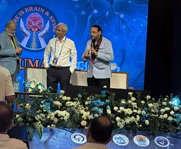
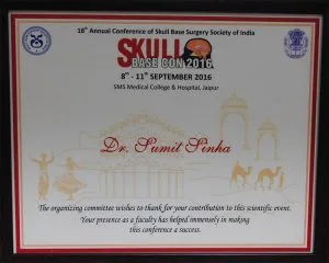
- Invited faculty at national skull base surgery conferences.
- Access to neuro-navigation, neuromonitoring, advanced imaging and multidisciplinary neuro-oncology care at Max Super Speciality Hospital, Dwarka, New Delhi.
Factors That Influence the Cost of Brain Tumor Surgery
- Type, size and location of the tumor.
- Surgical approach - endoscopic endonasal, keyhole craniotomy or conventional craniotomy.
- Use of navigation, neuromonitoring, awake mapping or special equipment.
- ICU and hospital stay.
- Further treatment such as radiation or chemotherapy.
- Insurance coverage - most policies cover medically necessary brain tumor surgery.
Meningioma - Why Location Matters
Meningiomas illustrate how the same type of tumor can pose very different challenges depending on where it grows. A meningioma over the surface of the brain (convexity meningioma) is usually straightforward to remove completely together with its dural attachment, and the chance of cure is high. Meningiomas along the midline falx or near the large venous sinuses require careful handling of these vital veins. Skull base meningiomas - around the olfactory groove, tuberculum sellae, sphenoid wing, cavernous sinus, petroclival region or foramen magnum - may surround arteries and cranial nerves controlling vision, eye movement, facial sensation, hearing and swallowing. In these locations, the goal is maximal safe removal while preserving function; small residual portions attached to critical structures may be monitored or treated with radiosurgery. Many small meningiomas discovered incidentally, especially in older people, never grow significantly and can simply be observed with periodic scans.
Gliomas and the Role of Molecular Diagnosis
Gliomas are now classified not only by how the cells look under the microscope but also by their molecular features - such as IDH mutation, 1p/19q co-deletion and MGMT promoter methylation. These markers give valuable information about prognosis and response to treatment. For example, IDH-mutant gliomas generally behave less aggressively than IDH-wild-type tumors, and MGMT methylation predicts better response to the chemotherapy drug temozolomide. Surgery aims for the maximum safe removal of the tumor, because evidence shows that a more complete resection is associated with better outcomes in many gliomas. Techniques such as awake mapping, tractography and fluorescence guidance help achieve this while protecting important functions. After surgery, the neuro-oncology team plans radiation and chemotherapy based on the tumor's grade and molecular profile.
Brain Metastases - A Changing Outlook
With improvements in cancer treatment, more patients live long enough for cancer to spread to the brain, and more patients with brain metastases are living well for extended periods. Treatment depends on the number, size and location of the metastases, the control of cancer elsewhere and the patient's overall condition. A single large metastasis causing pressure symptoms is often best removed surgically, followed by focused radiation to the cavity. Multiple small metastases may be treated with stereotactic radiosurgery. Newer targeted therapies and immunotherapies can also be effective against brain metastases from certain cancers. Decisions are made jointly by the neurosurgeon, radiation oncologist and medical oncologist.
Seizures, Driving and Daily Life
Seizures are a common symptom of brain tumors, particularly those involving the cerebral cortex. Anti-seizure medicines are prescribed when seizures have occurred and, in some cases, around the time of surgery. After tumor removal, many patients experience fewer or no seizures, and medicines may be reduced over time under medical supervision. Driving restrictions apply after a seizure and after some brain operations; the duration depends on the type of surgery and seizure history, and patients should follow their doctor's advice and local regulations. Most people are able to return to work, family life and hobbies after recovery, with adjustments as needed during any additional treatment.
Rebuilding the Skull Base After Endonasal Surgery
When a tumour is removed through the nose, the surgeon creates an opening in the thin bone and lining that separate the nasal cavity from the brain. Closing this opening securely is as important as removing the tumour, because a persistent gap can allow cerebrospinal fluid to leak into the nose and, more seriously, allow infection to reach the brain. The method of repair is planned before surgery according to the size of the tumour and how much the fluid spaces around the brain are likely to be opened during removal.
How the repair is done
- Layered closure: small defects are sealed with layers of fat, fascia or graft material and tissue sealants.
- Vascularised nasal flap: for larger openings, a flap of the nose's own lining, kept attached to its blood supply, is rotated to cover the defect. Because it has a living blood supply, it heals reliably.
- Nasal packing or splints: sometimes placed for a few days to support the repair.
- Lumbar drain: occasionally used for a short period to reduce fluid pressure while the repair heals.
After surgery, watery, salty-tasting fluid dripping from the nose - especially on bending forward - should be reported immediately, as it may indicate a leak that needs attention.
When a Biopsy Is the First Step
Not every brain tumour should be removed as the first step. Some lesions lie deep in the brain, in areas where removal would cause unacceptable harm. Others, such as primary brain lymphoma or certain germ cell tumours, are best treated with chemotherapy or radiotherapy rather than surgery, and some lesions that look like tumours on MRI turn out to be infections, such as tuberculomas, or inflammatory conditions. In these situations, obtaining a small sample of tissue to establish an accurate diagnosis is the most valuable thing surgery can offer.
A stereotactic biopsy uses navigation to guide a fine needle to the target through a small burr hole with high precision. For lesions within or next to the ventricles, an endoscopic biopsy allows the surgeon to see the tumour directly, and associated hydrocephalus can be treated in the same sitting. Recovery is usually quick, and the results guide the neuro-oncology team in planning the most appropriate treatment.
Questions Worth Asking Before Surgery
A consultation is a two-way conversation, and patients who understand their plan tend to feel more in control. Writing questions down beforehand helps. Useful questions include: What type of tumour is this likely to be, and how certain is that? What is the goal of surgery - complete removal, partial removal or biopsy? Which approach is planned, and why is it suitable for this tumour? What are the specific risks in this location? Will further treatment be needed? How long will recovery take, and when can I return to work and driving?
Patients from outside Delhi can send their MRI and reports by WhatsApp on +91-8448877746 for a preliminary opinion, and video consultations are available. To meet Dr. Snha at Max Super Speciality Hospital, Dwarka, from Monday to Saturday, book an appointment. Related information is available on our hydrocephalus treatment page.
Frequently Asked Questions
Are all brain tumors cancerous?
No. Many brain tumors, such as most meningiomas, pituitary adenomas and schwannomas, are benign. However, even benign tumors may need treatment because they can press on important brain structures.
Can a brain tumor be removed through the nose?
Yes. Pituitary tumors and many skull base tumors can be removed through the nose using an endoscope, without any external incision.
Is keyhole brain surgery as effective as conventional surgery?
For suitable tumors, keyhole and endoscopic approaches can achieve similar tumor removal with less trauma to healthy tissue. The choice depends on the tumor's size, location and nature.
How long does it take to recover from brain tumor surgery?
Many patients go home within a few days after minimally invasive surgery and resume light activities in a few weeks. Recovery varies with the tumor type and any additional treatment.
Will I need radiation after surgery?
It depends on the final pathology and how completely the tumor was removed. Many completely removed benign tumors need no further treatment.
Can a brain tumor come back?
Some tumors can recur, which is why follow-up MRI scans are important. Recurrence depends on the tumor type, grade and extent of removal.
Is brain surgery safe in elderly patients?
With careful assessment and minimally invasive techniques, many elderly patients undergo brain tumor surgery safely. The decision considers overall health and the expected benefit.
Expert Brain Tumor Care in Delhi
If you or a loved one has been diagnosed with a brain tumor, or has symptoms such as persistent headaches, seizures, vision changes or weakness, an early expert opinion is valuable. Book a consultation with Dr. (Prof.) Sumiet Snha at Max Hospital, Dwarka, New Delhi. Related pages: acoustic neuroma, hydrocephalus and vascular neurosurgery.