Hearing the words "tumor in the spine" is frightening for any patient and family. Many immediately assume the worst - cancer, paralysis, a lifetime of disability. In reality, spinal tumors are a diverse group of conditions. A large number are benign (non-cancerous) and can be removed completely with excellent results, while many malignant tumors can be controlled effectively with a combination of surgery, radiation and medical treatment. What matters most is an accurate diagnosis, a clear plan and a surgical team with deep experience in spinal cord and nerve surgery.
Dr. (Prof.) Sumiet Snha, former Professor of Neurosurgery at AIIMS, New Delhi, and Principal Director - Neurosurgery & Spine Surgery at Max Hospital, Dwarka, treats the full spectrum of spinal tumors - from small nerve sheath tumors removed through keyhole incisions to complex vertebral tumors requiring reconstruction of the spine. He has led marathon multi-specialty operations for rare conditions such as en-bloc removal of a lumbar chordoma, performed together with oncology, cardiothoracic and urology teams. This page explains the types of spinal tumors, their symptoms, how they are diagnosed and the modern surgical options available.
What Is a Spinal Tumor?
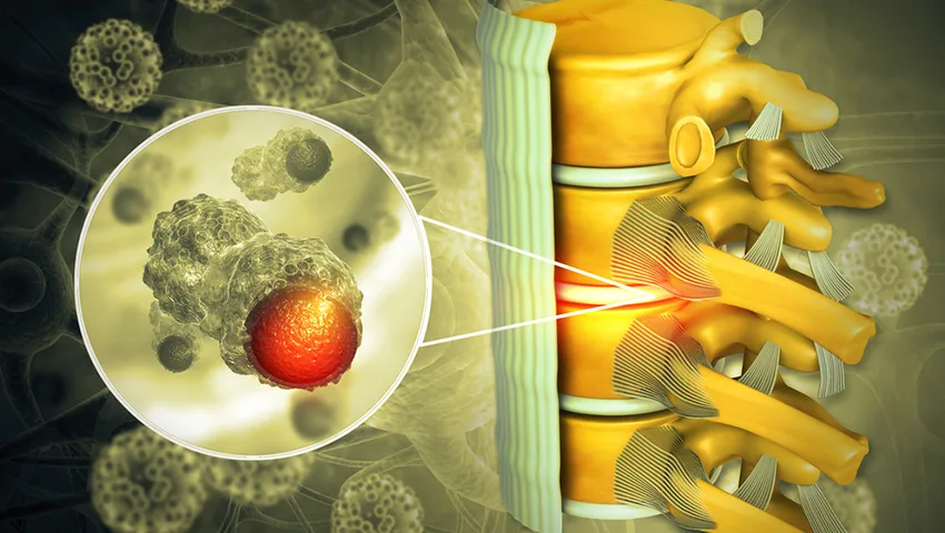
A spinal tumor is an abnormal growth of cells within or around the spinal column - the vertebrae, the spinal cord, its coverings (meninges) or the nerve roots. Tumors cause problems in two main ways: by pressing on the spinal cord or nerves, leading to pain, numbness and weakness, and by weakening the bone, which can lead to fractures and instability. Malignant tumors may also spread to other parts of the body.
Spinal tumors are classified by their location relative to the spinal cord and its covering, the dura. This classification guides both diagnosis and surgical approach.
Extradural tumors
Outside the dura, usually arising from the vertebral bones. The most common are metastases (spread from cancers elsewhere), along with myeloma, lymphoma, haemangioma, chordoma and bone tumors.
Intradural-extramedullary tumors
Inside the dura but outside the spinal cord. Mostly benign - meningiomas, schwannomas and neurofibromas - and often completely removable.
Intramedullary tumors
Within the substance of the spinal cord itself - ependymomas, astrocytomas and haemangioblastomas. These need the most delicate microsurgery.
Dumbbell (foraminal) tumors
Nerve sheath tumors that grow both inside the canal and outwards through the foramen, sometimes into the chest or abdomen.
Primary vs Secondary (Metastatic) Spine Tumors
Primary spinal tumors begin in the spine itself. They are relatively uncommon and include benign lesions like meningioma, schwannoma, osteoid osteoma and haemangioma, as well as malignant ones like chordoma, chondrosarcoma and Ewing's sarcoma.
Secondary or metastatic tumors are far more common. The spine is one of the most frequent sites to which cancers spread, particularly cancers of the breast, lung, prostate, kidney and thyroid, as well as multiple myeloma. In some patients, back pain from a spinal metastasis is the very first sign of an undiagnosed cancer. Advances in cancer care mean that many patients with spinal metastases live for years, making it important to protect their mobility, independence and comfort.
Common Spinal Tumor Types Explained
Spinal meningioma
Meningiomas arise from the meninges, the thin membranes covering the spinal cord. They are among the most common intradural tumors in adults and are seen more often in women, frequently in the thoracic (mid-back) region. They grow slowly, so symptoms such as leg stiffness, numbness and a gradual change in walking may develop over months or years and are sometimes mistaken for arthritis or ageing. Because they sit outside the cord, they can usually be removed completely with microsurgery, along with the small area of dura to which they are attached, and the long-term results are generally excellent.
Schwannoma and neurofibroma
These nerve sheath tumors grow from the insulating layer of a spinal nerve root. Schwannomas are typically solitary and benign, and often cause radicular pain along the course of the affected nerve - for example, a band of pain around the chest or sciatica-like pain in the leg. Some extend through the foramen in a "dumbbell" shape. Neurofibromas are more closely intertwined with the nerve fibres and are associated with neurofibromatosis type 1. Many schwannomas are ideally suited to minimally invasive removal with nerve-function monitoring.
Ependymoma
Ependymomas are the most common tumors within the spinal cord in adults. They often have a relatively clear boundary with normal cord tissue, which allows gross total removal in experienced hands. A specific variant, the myxopapillary ependymoma, arises at the lower end of the cord (the filum terminale) and may present with long-standing back pain and sciatica.
Astrocytoma
Astrocytomas infiltrate the spinal cord tissue and are more common in children. Because they lack a clear plane, surgery aims to remove as much tumor as can be safely achieved, and additional therapy may be required depending on the grade.
Haemangioma
Vertebral haemangiomas are very common benign vascular lesions, usually discovered incidentally on MRI and needing no treatment. Rarely, an aggressive haemangioma expands the bone and compresses the cord, requiring embolisation, cement augmentation, surgery or radiation.
Metastases and multiple myeloma
Metastatic tumors from breast, lung, prostate, kidney and thyroid cancers most often involve the vertebral body. Multiple myeloma, a cancer of plasma cells, frequently causes painful vertebral fractures. Treatment combines oncological therapy with radiation and, when needed, surgery or cement augmentation to relieve pain, prevent collapse and protect the spinal cord.
Chordoma and other primary bone tumors
Chordomas arise from remnants of embryonic tissue, typically at the base of the skull or the sacrum and occasionally in the vertebrae. They grow slowly but are locally aggressive, and the best long-term control is achieved with complete (en-bloc) removal, often followed by specialised radiation.
Symptoms of Spinal Tumors
Symptoms depend on the tumor's location, size and speed of growth. Common warning signs include:
- Back or neck pain that is persistent, progressive and often worse at night or when lying down, not relieved by rest.
- Pain radiating into the arms, around the chest or abdomen (band-like), or into the legs.
- Numbness, tingling or reduced sensitivity to temperature and touch.
- Weakness in the arms or legs, difficulty walking, frequent falls or a feeling of stiffness in the legs.
- Loss of fine hand function - dropping objects, difficulty writing or buttoning.
- Bladder or bowel dysfunction - urgency, retention or incontinence.
- Scoliosis or a new spinal deformity, particularly in children.
- Unexplained weight loss, fever or fatigue with back pain.
Red flags - seek prompt medical attention
Back pain in a person with a known history of cancer, new weakness or numbness in the legs, difficulty passing urine or rapidly increasing pain should never be ignored. Spinal cord compression from a tumor is a medical emergency - the sooner pressure is relieved, the better the chance of preserving or recovering the ability to walk.
How Spinal Tumors Are Diagnosed
- Clinical evaluation - history (including any previous cancer), detailed neurological examination and assessment of pain pattern.
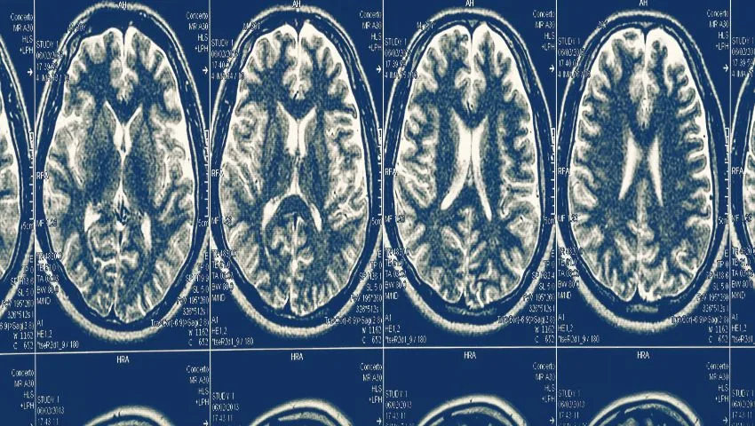
- MRI with contrast - the most important test, showing the tumor's location, its relation to the spinal cord and nerves, and features suggesting its type.
- CT scan - to assess bone destruction, calcification and stability, and for surgical planning of screw placement.
- PET-CT or whole-body imaging - when metastatic disease is suspected, to find the primary cancer and other sites.
- Blood tests - including tests for myeloma and tumor markers where appropriate.
- Biopsy - a CT-guided needle biopsy or surgical biopsy to confirm the exact diagnosis when it influences treatment.
- Stability assessment - scoring systems help decide whether the spine is at risk of collapse and needs stabilisation.
Treatment Options for Spinal Tumors
Treatment is tailored to the tumor type, its location, the patient's neurological status, general health and, in the case of cancer, the overall disease picture. Many patients are best managed by a multidisciplinary team including the neurosurgeon, medical oncologist, radiation oncologist, radiologist, pathologist and rehabilitation specialists.
Observation
Small, asymptomatic benign tumors (e.g. some haemangiomas) may simply be monitored with periodic MRI.
Surgery
To remove the tumor, relieve cord/nerve compression, obtain tissue for diagnosis and stabilise the spine.
Radiation therapy
Conventional radiotherapy or high-precision stereotactic radiosurgery (SBRT) - often after surgery for malignant tumors.
Medical treatment
Chemotherapy, targeted therapy, immunotherapy, hormone therapy and bone-strengthening medicines, as appropriate for the cancer type.
Surgical Approaches - From Keyhole to Complex Reconstruction
Minimally invasive and endoscopic tumor removal
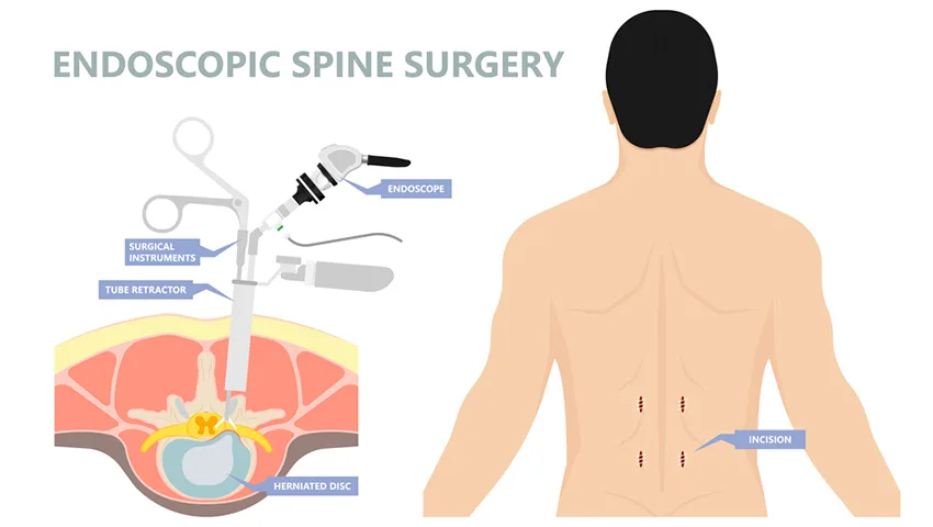
Many intradural-extramedullary tumors such as schwannomas and meningiomas, particularly when small to medium in size, can be removed through a small midline or paramedian incision using tubular retractors, the operating microscope or the endoscope. Only a limited amount of bone is removed (hemilaminectomy), preserving the stabilising structures of the spine. The dura is opened, the tumor is dissected away from the spinal cord and nerve roots under high magnification, and the dura is closed watertight. Patients typically mobilise early and have less post-operative pain than after traditional open surgery.
Microsurgical removal of intramedullary tumors
Tumors within the spinal cord require meticulous microsurgery. The cord is opened in the midline and the tumor is removed while preserving healthy neural tissue. Intra-operative neuromonitoring (recording the signals travelling through the spinal cord) is used to guide the extent of removal and protect function.
Separation surgery for metastatic disease
When cancer in the vertebra is compressing the spinal cord, "separation surgery" removes the tumor that is pressing on the cord and creates a safety margin. The spine is stabilised with screws and rods, often placed percutaneously through small incisions. High-dose precision radiation can then be delivered safely to the remaining tumor.
Vertebroplasty and kyphoplasty
For painful vertebral fractures caused by tumors without significant cord compression, bone cement can be injected into the vertebra through a needle to relieve pain and prevent further collapse.
En-bloc resection
Certain primary malignant tumors like chordoma and some sarcomas are best treated by removing the tumor-bearing vertebra in one piece with a margin of healthy tissue, followed by reconstruction of the spine with cages and instrumentation. These are among the most complex operations in spine surgery, requiring careful planning and a multi-specialty team.
Technology That Improves Safety
- Intra-operative neuromonitoring - continuous monitoring of motor and sensory pathways during surgery.
- High-definition operating microscope and endoscope - for precise dissection of tumors from delicate neural tissue.
- Neuro-navigation and intra-operative imaging - for accurate localisation and screw placement.
- Ultrasonic aspirators - to debulk tumors gently from inside.
- Minimally invasive instrumentation - percutaneous screws that stabilise the spine with minimal muscle damage.
A Case That Illustrates Complex Spine Tumor Care
A 46-year-old woman had severe lower back pain for two months, limiting her daily life. Investigations revealed a chordoma - a rare malignant tumor - in the L2 vertebral body. To give her the best chance of cure, the plan was to remove the entire vertebral body in a single piece containing the tumor, avoiding spillage of tumor cells. A team including neurosurgery led by Prof. Sumiet Snha, surgical oncology, cardiothoracic and urology surgeons carried out a staged operation - first from the back and then from the front - on the same day, lasting around 14 hours. The surgery was successful; her symptoms resolved and she returned home pain-free and resumed her normal activities. Such operations are performed at very few centres, and they demonstrate the importance of experience, planning and teamwork in spinal oncology.
Radiation Therapy and Radiosurgery for Spinal Tumors
Radiation plays an important role in the treatment of many spinal tumors, either on its own or after surgery. Conventional external beam radiotherapy delivers treatment over several sessions and is effective for radiosensitive tumors such as lymphoma, myeloma and some metastases. Stereotactic body radiotherapy (SBRT), also called spinal radiosurgery, uses highly precise, image-guided beams to deliver high doses to the tumor while sparing the nearby spinal cord. It has made it possible to control tumors that were previously considered "radioresistant", such as those from kidney cancer or melanoma.
The surgeon and the radiation oncologist plan together. When the spinal cord is already compressed, surgery usually comes first to create a safe gap (the separation surgery described above), after which radiosurgery treats the remaining disease. When there is no compression or instability, radiation alone may be the best option. For benign tumors that cannot be removed completely because of their location, radiosurgery can help control growth. Side effects are generally mild and temporary, such as fatigue or skin irritation, and are monitored closely.
Nutrition, Bone Health and General Wellbeing
Good nutrition supports wound healing and recovery. A diet rich in protein, fruits and vegetables, along with adequate hydration, is recommended before and after surgery. Patients with cancer may need a dietitian's advice to maintain weight during treatment. Bone health is especially important for those with metastatic disease or osteoporosis: vitamin D, calcium and bone-strengthening medicines prescribed by the oncologist or physician reduce the risk of fractures. Gentle, regular activity as advised by the physiotherapist helps maintain muscle strength, balance and mood.
Risks of Spinal Tumor Surgery
All spinal tumor surgery carries some risk, which depends heavily on the tumor type and location. Possible complications include temporary or permanent worsening of neurological function, cerebrospinal fluid leak, infection, bleeding, wound problems, instability and, for malignant tumors, recurrence. In most benign intradural tumors, the risk of lasting deficit is low and the chance of complete removal and cure is high. Your surgeon will explain the expected benefits and specific risks in your case so that you can make an informed decision.
Recovery and Rehabilitation
What to expect
- Hospital stay: usually a few days for minimally invasive removal of benign tumors; longer for complex reconstruction.
- Early mobilisation: walking with physiotherapy support begins as soon as it is safe, often within a day or two.
- Neurological recovery: numbness and weakness present before surgery may continue to improve over weeks to months.
- Pathology report: the final tissue diagnosis guides whether further treatment such as radiation is needed.
- Follow-up MRI: scheduled scans to confirm removal and monitor for recurrence.
Rehabilitation plays a vital role. Physiotherapy improves strength, balance and walking; occupational therapy helps with hand function and daily activities; and bladder training may be needed if bladder function was affected. Emotional support and counselling are equally important, particularly for patients dealing with a cancer diagnosis.
Spinal Tumors in Children
Although uncommon, spinal tumors can occur in children and may present with back pain, a new curvature of the spine (scoliosis), a change in walking pattern, clumsiness or new bladder problems. Children with persistent night-time back pain or neurological symptoms should always be evaluated. Paediatric spinal surgery requires special attention to the growing spine to prevent deformity later in life.
Why Choose Dr. (Prof.) Sumiet Snha?
- AIIMS-trained neurosurgeon - MBBS, MS, DNB, MCh (AIIMS, New Delhi), FACS - with over 25 years of experience.
- Former Professor of Neurosurgery at AIIMS, New Delhi, with extensive experience in complex spinal cord and spinal column tumors.
- Expertise in minimally invasive, endoscopic and microsurgical tumor removal as well as complex reconstruction and en-bloc resection.
- Close collaboration with oncology, radiation and rehabilitation teams for comprehensive cancer care.
- Advanced infrastructure at Max Super Speciality Hospital, Dwarka, including neuromonitoring, navigation and high-end imaging.
- Compassionate, transparent communication with patients and families at every step.
Factors Affecting the Cost of Spinal Tumor Surgery
- Type, size and location of the tumor.
- Need for stabilisation with screws, rods or cages.
- Use of neuromonitoring, navigation and special equipment.
- Duration of ICU and hospital stay.
- Further treatments such as radiation or chemotherapy.
- Insurance coverage and hospital room category.
Living With and After a Spinal Tumor
For most patients with benign tumors, successful surgery is the end of the problem, followed by periodic MRI checks. For patients with cancer that has spread to the spine, the aim is to control pain, maintain or restore the ability to walk, prevent fractures and support ongoing oncological treatment. Regular exercise within safe limits, good nutrition, bone health measures and emotional support all contribute to a better quality of life. Families are encouraged to be involved in planning and rehabilitation, and patients should report any new pain, weakness or bladder change promptly.
Questions to Ask Your Surgeon
Being well informed helps you feel more in control. Consider asking your surgeon:
- What type of tumor is it likely to be, and how certain is the diagnosis before surgery?
- Can the tumor be removed completely, and what happens if a small part is left behind?
- Is a minimally invasive or endoscopic approach suitable in my case?
- Will I need screws and rods to stabilise my spine?
- What are the specific risks to my walking, sensation, and bladder or bowel function?
- Will I need radiation or other treatment afterwards?
- How long will I be in hospital and how long before I can return to work?
- How often will I need follow-up MRI scans?
Dr. Snha encourages patients and families to bring their questions written down. Taking time to understand the plan leads to better decisions and a smoother recovery.
The Value of a Second Opinion
Spinal tumor surgery is a major decision, and it is entirely reasonable to seek a second opinion before proceeding. A second review may confirm the diagnosis, suggest a less invasive approach, clarify whether surgery is needed urgently or can be planned, or highlight additional investigations that would improve safety. Patients from across India and abroad share their MRI scans, CT images and reports with Dr. Snha for an expert opinion. When there is spinal cord compression with worsening weakness, however, delays can be harmful - in such cases the priority is prompt decompression.
Supporting the Whole Patient
A spinal tumor affects much more than the spine. Patients may face pain, anxiety about the future, financial concerns and changes in their role at work or at home. Good care addresses all of these. Pain is managed with a combination of medicines, positioning and, where appropriate, interventional techniques. Physiotherapists and occupational therapists help patients regain independence. For those with cancer, palliative and supportive care specialists work alongside the surgical and oncology teams from an early stage - not only at the end of life - to control symptoms and improve quality of life. Families receive clear information about what to expect and how to help at home, including safe transfers, skin care for those with limited mobility and early signs that need medical attention.
Warning Signs After Surgery
Most patients recover smoothly, but it is important to know when to contact the team. Call the hospital or seek urgent care if you notice fever, increasing redness, swelling or discharge from the wound, clear fluid leaking from the incision, a severe headache that worsens on sitting up (which may indicate a spinal fluid leak), new or worsening weakness or numbness, difficulty passing urine, or pain in the calf with swelling (a possible sign of a blood clot). Early attention to these issues allows quick and effective treatment.
How Surgeons Assess Spinal Stability in Cancer
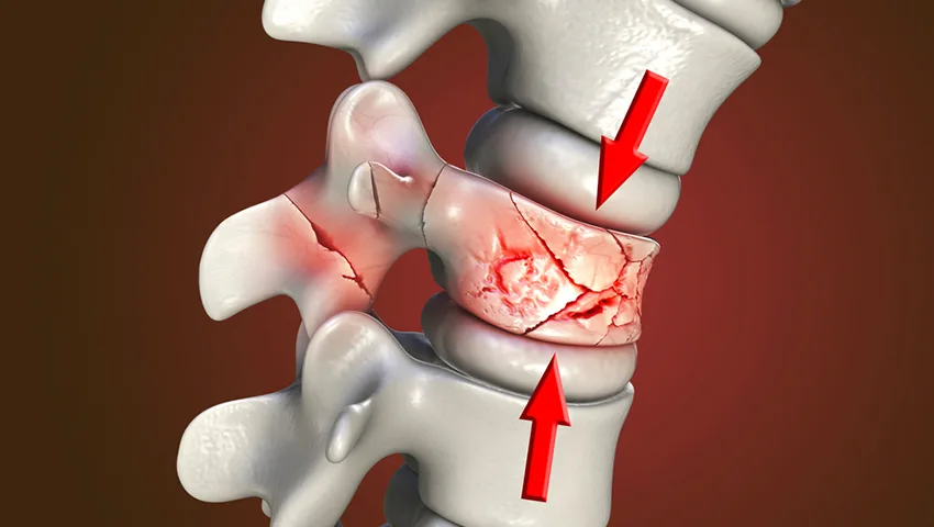
When cancer spreads to the spine, two separate questions shape treatment: is the spinal cord or nerve being compressed, and is the spine still mechanically stable? A vertebra weakened by tumour may collapse or slip even without cord compression, producing severe pain on movement and a risk of sudden neurological injury. Recognising instability early allows it to be treated in a planned way - often with minimally invasive fixation or cement augmentation - before a collapse turns a manageable problem into an emergency.
Features that suggest instability
- Mechanical pain: pain that worsens sharply on sitting up, standing or turning, and eases on lying still.
- Location: junctional regions such as the neck-chest (cervicothoracic) and chest-lumbar (thoracolumbar) junctions are more vulnerable.
- Type of bone destruction: lytic (bone-dissolving) lesions weaken the vertebra more than sclerotic (bone-forming) ones.
- Collapse and alignment: loss of vertebral height, new angulation or slippage on imaging.
- Involvement of the back elements: tumour affecting the pedicles and facet joints on one or both sides.
These features are combined into a structured score, such as the Spinal Instability Neoplastic Score (SINS), which helps the surgical, radiation and medical oncology teams agree on whether stabilisation is needed before or alongside radiotherapy. A stable spine without compression can often be treated with radiation alone, whereas an unstable one generally needs mechanical support first.
Coordinating Surgery With Cancer Treatment
Spine surgery for cancer is one step in a longer treatment plan. Timing matters: radiotherapy is usually delayed for a short period after surgery to allow the wound to heal, and some chemotherapy or targeted medicines may be paused around the operation. Smaller incisions and minimally invasive fixation help patients recover quickly so that cancer treatment can resume with minimal interruption. Dr. Snha works closely with the treating oncologists to agree a timeline, and patients are given a clear written summary of what happens next and whom to contact at each stage.
Frequently Asked Questions
Are all spinal tumors cancerous?
No. Many spinal tumors, especially those inside the dura but outside the spinal cord such as meningiomas and schwannomas, are benign and can often be removed completely.
Can spinal tumors be removed through keyhole surgery?
Many small and medium-sized benign tumors can be removed through minimally invasive or endoscopic approaches. Larger or more complex tumors may need open surgery or reconstruction.
Will I be paralysed after spinal tumor surgery?
The aim of surgery is to protect and restore nerve function. With modern microsurgery and neuromonitoring the risk of serious new deficit is low for most benign tumors, though it varies with the tumor type and location.
Why does spinal tumor pain get worse at night?
Tumor-related pain is often persistent and may worsen when lying down, partly due to changes in pressure and the absence of daytime distraction. Night pain is a warning sign that deserves evaluation.
Do I need radiation after surgery?
It depends on the final pathology. Benign tumors that are completely removed usually need no further treatment. Malignant or incompletely removed tumors may need radiation or other therapy.
Can a spinal tumor come back?
Recurrence depends on the tumor type and the extent of removal. Regular follow-up MRI scans help detect any regrowth early.
What is the recovery time after spinal tumor surgery?
After minimally invasive removal of a benign tumor, many patients go home within a few days and return to light activities in a few weeks. Complex surgeries require longer recovery and rehabilitation.
Expert Care When It Matters Most
If you or a loved one has been diagnosed with a spinal tumor, or has symptoms that raise concern, early expert assessment can make a real difference. Book a consultation with Dr. (Prof.) Sumiet Snha at Max Hospital, Dwarka, New Delhi. You can also read about nerve sheath tumors, brain tumor surgery and spinal fractures.