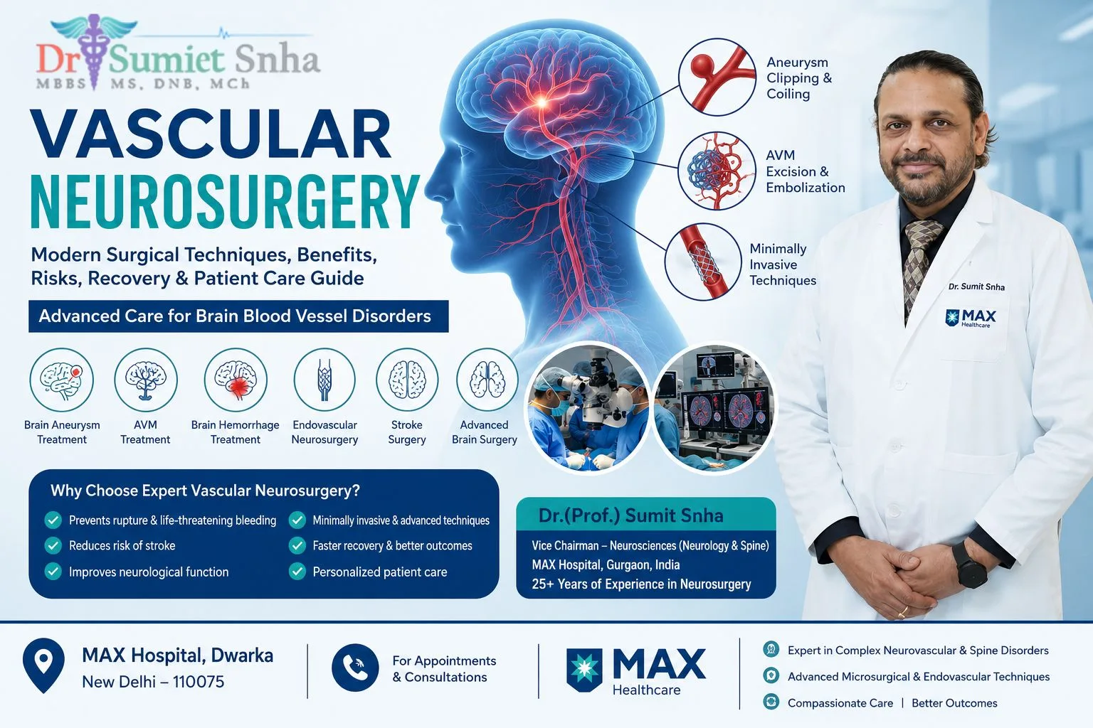
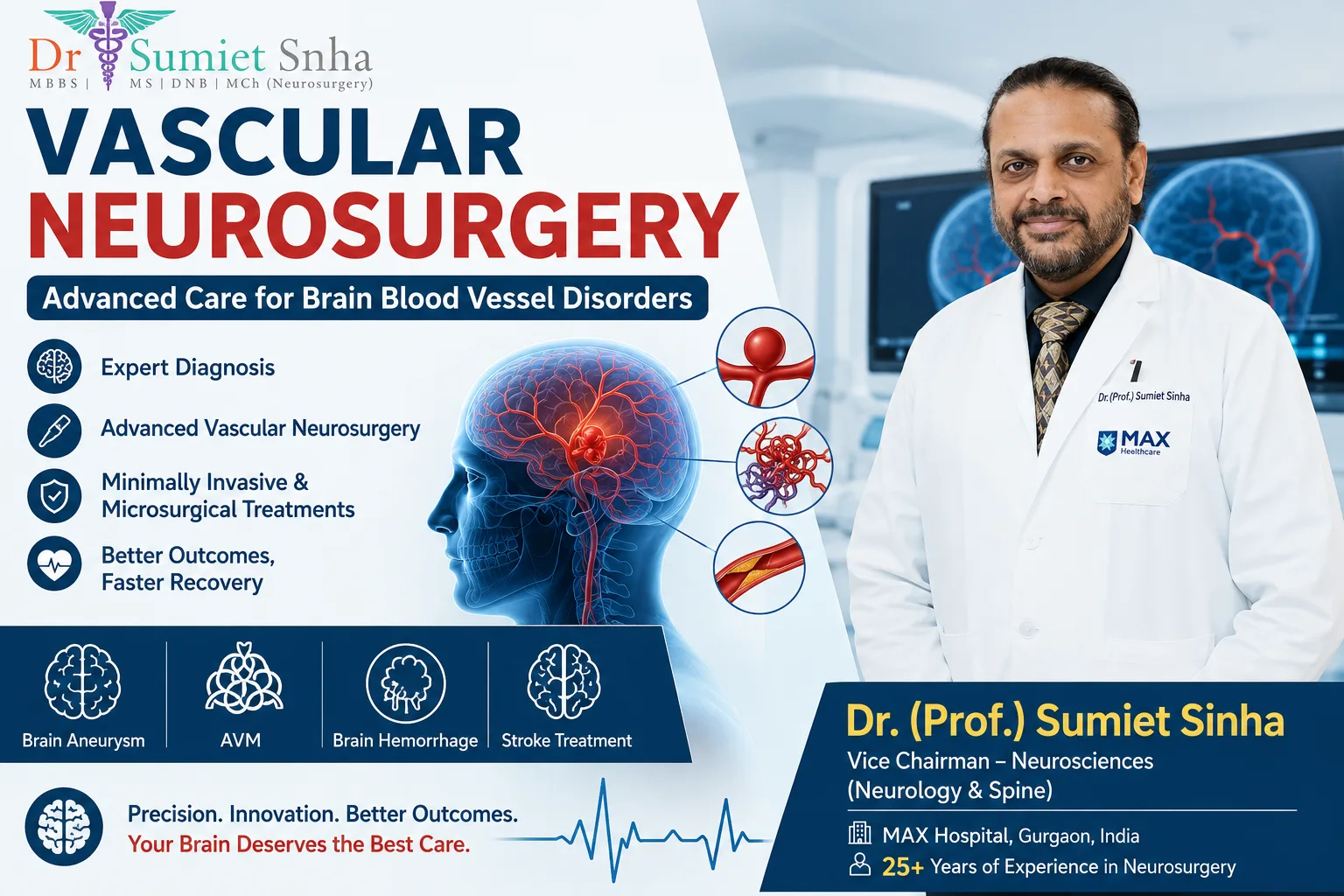
The brain receives about one-fifth of the body's blood supply through an intricate network of arteries and veins. When these vessels develop abnormalities - a weak, ballooning spot in an artery wall, a tangle of abnormal vessels, or a blockage - the consequences can be sudden and serious: a bleed in the brain, a stroke, seizures or disabling headaches. Vascular neurosurgery (cerebrovascular neurosurgery) is the specialty that diagnoses and treats these conditions, combining delicate microsurgery with modern endovascular techniques and intensive neuro-critical care.
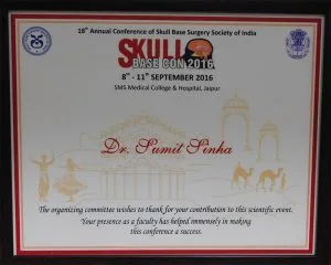
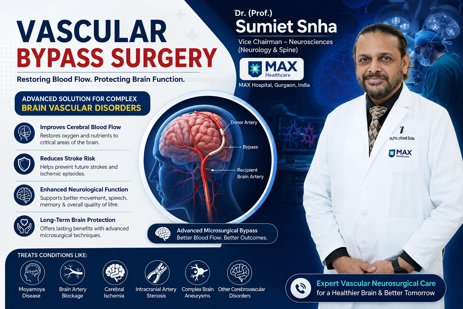
Dr. (Prof.) Sumiet Snha, former Professor of Neurosurgery at AIIMS, New Delhi, has a long-standing interest in cerebrovascular surgery. He was awarded the Young Neurosurgeon Award to attend the 9th International Conference on Cerebrovascular Surgery in Nagoya, Japan, and trained with renowned cerebrovascular surgeon Prof. Hiroshi Sano of Japan. At Max Hospital, Dwarka, New Delhi, he treats complex arteriovenous malformations (AVMs), aneurysms and other vascular lesions of the brain and spinal cord. This page explains these conditions and their treatment in detail.
Conditions Treated in Vascular Neurosurgery
Brain aneurysm
A weak, bulging spot on an artery wall that can rupture and cause a life-threatening bleed (subarachnoid haemorrhage).
Arteriovenous malformation (AVM)
An abnormal tangle of arteries and veins without the normal capillary network, prone to bleeding and seizures.
Cavernous malformation (cavernoma)
A cluster of abnormal, thin-walled vessels resembling a mulberry, which can cause seizures or small bleeds.
Dural arteriovenous fistula
An abnormal connection between arteries and veins in the covering of the brain, which may cause pulsatile tinnitus or bleeding.
Intracerebral haemorrhage
Bleeding within the brain, often due to high blood pressure; large clots may need surgical removal.
Moyamoya and occlusive disease
Narrowing or blockage of major brain arteries, sometimes treated with bypass surgery - see our vascular bypass page.
Carotid artery disease
Narrowing of the neck arteries that can cause stroke; treated with medicines, endarterectomy or stenting.
Spinal vascular malformations
AVMs and fistulas of the spinal cord that can cause progressive weakness or sudden paralysis.
Brain Aneurysms
What is a brain aneurysm?
A brain (cerebral) aneurysm is a balloon-like bulge that develops at a weak point in an artery wall, most commonly at branching points of the arteries at the base of the brain (the circle of Willis). Many aneurysms are small and never cause problems; they may be discovered incidentally on a scan done for another reason. However, if an aneurysm ruptures, blood spills into the space surrounding the brain - a subarachnoid haemorrhage (SAH) - a medical emergency with significant risk of death or disability.
Risk factors
- High blood pressure.
- Smoking - one of the strongest modifiable risk factors.
- Family history of aneurysms (two or more first-degree relatives).
- Certain inherited conditions, such as polycystic kidney disease and connective tissue disorders.
- Female sex and increasing age.
- Heavy alcohol use and stimulant drug use.
Symptoms
Unruptured aneurysms are usually silent, although large ones may press on nerves, causing a drooping eyelid, double vision, a dilated pupil or facial pain. A ruptured aneurysm typically causes:
- A sudden, extremely severe headache - often described as "the worst headache of my life" or a "thunderclap" headache reaching full intensity within seconds.
- Nausea and vomiting.
- Neck stiffness and sensitivity to light.
- Loss of consciousness, confusion or seizures.
A sudden "thunderclap" headache is an emergency
Call emergency services or go to the nearest hospital with neurosurgical facilities immediately. Early diagnosis with a CT scan and prompt treatment of a ruptured aneurysm save lives and reduce disability. Some people experience a smaller "warning leak" headache days before a major bleed - this also needs urgent evaluation.
Diagnosis
A plain CT scan of the head detects most subarachnoid haemorrhages in the first hours. CT angiography shows the aneurysm's size, shape and location. Digital subtraction angiography (DSA) - a catheter-based study - provides the most detailed images and allows endovascular treatment in the same session when appropriate. MRI and MR angiography are useful for screening and follow-up.
Treatment options
The goal is to seal off the aneurysm from the circulation to prevent it from bleeding (or re-bleeding). The two main techniques are:
Microsurgical clipping
Through a craniotomy, the surgeon places a tiny titanium clip across the neck of the aneurysm under the microscope, excluding it from the circulation permanently while preserving the parent artery.
Endovascular coiling / flow diversion
Through a catheter from the groin or wrist, the aneurysm is packed with coils, or a flow-diverting stent is placed across it, promoting clotting and healing.
The choice depends on the aneurysm's location, shape, size and neck, the patient's age and condition, and whether there is a large blood clot needing removal. Many centres, including our team, make this decision collaboratively between the vascular neurosurgeon and the interventional neuroradiologist to offer each patient the best option. Unruptured aneurysms are evaluated individually - some are best treated, others are safely monitored with blood pressure control and smoking cessation.
Care After Subarachnoid Haemorrhage
Securing a ruptured aneurysm is only the first step. Patients then need careful monitoring in a neuro-intensive care unit for about two weeks, because several complications can arise:
- Vasospasm and delayed cerebral ischaemia - narrowing of brain arteries typically between days 3 and 14, which can cause stroke-like symptoms. It is prevented and treated with medicines such as nimodipine, careful fluid management and, if needed, endovascular therapy.
- Hydrocephalus - blood can block the flow of cerebrospinal fluid, requiring a temporary drain (external ventricular drain) or, later, a shunt.
- Seizures, sodium imbalance, fever and heart strain - monitored and treated promptly.
Recovery after subarachnoid haemorrhage varies widely. Many patients who are in good condition when treated make a good recovery, though fatigue, memory and concentration difficulties, headaches and mood changes are common in the months afterwards. Neuro-rehabilitation and patience are key.
Arteriovenous Malformations (AVMs)
What is an AVM?
Normally, blood flows from arteries into a fine network of capillaries, where oxygen is delivered to tissues, before draining into veins. In an arteriovenous malformation, arteries connect directly to veins through a tangle of abnormal vessels called the nidus, bypassing the capillaries. The high-pressure flow can make the vessels fragile and prone to rupture, and can "steal" blood from surrounding brain. Most AVMs are thought to be present from birth, although they often cause symptoms only in young adulthood.
Symptoms
- Brain haemorrhage - sudden headache, vomiting, weakness, speech problems or loss of consciousness.
- Seizures - often the first sign.
- Headaches, sometimes migraine-like, on the same side.
- Progressive neurological deficits from steal phenomenon or mass effect.
- A whooshing sound in the head (bruit) in some cases.
Grading and treatment decisions
AVMs are graded, commonly with the Spetzler-Martin scale, based on size, location in or near "eloquent" brain (areas controlling movement, speech, vision and sensation) and the pattern of venous drainage. The grade helps estimate surgical risk and guides the choice of treatment:
Microsurgical resection
Complete removal of the AVM through a craniotomy offers an immediate cure. Often preferred for lower-grade, accessible AVMs, especially after a bleed.
Endovascular embolisation
Glue-like materials are injected through catheters to block feeding vessels, usually to make surgery safer or as part of a combined strategy.
Stereotactic radiosurgery
Focused radiation for small, deep or eloquent-area AVMs, gradually causing the vessels to close over 1-3 years.
Observation
Some unruptured, high-grade AVMs may be safer to observe, with blood pressure control and seizure management.
Dr. Snha has special interest and experience in the surgical treatment of complex arteriovenous malformations, planning each case in detail with angiography, advanced MRI and, where needed, pre-operative embolisation.
Cavernous Malformations (Cavernomas)
Cavernomas are clusters of dilated, thin-walled blood vessels with slow blood flow. They may be single or multiple and can run in families. Many are discovered incidentally and never cause problems. Others cause seizures or small, repeated bleeds leading to headaches and neurological symptoms. MRI - particularly susceptibility-weighted sequences - is the best test to detect them; they are usually invisible on angiography. Surgical removal is considered for cavernomas that cause seizures resistant to medicines or that have bled and are accessible, including selected cavernomas in the brainstem, which require highly specialised microsurgery. Asymptomatic cavernomas are generally observed.
Brain Haemorrhage and Stroke Surgery
High blood pressure is the most common cause of bleeding within the brain (intracerebral haemorrhage). Small bleeds are managed with blood pressure control and supportive care. Large bleeds causing pressure, particularly in the cerebellum, may need surgical removal of the clot, sometimes through minimally invasive or endoscopic techniques. In severe strokes causing life-threatening brain swelling ("malignant" middle cerebral artery infarction), a decompressive craniectomy - temporarily removing part of the skull to allow the brain to swell - can be lifesaving. For acute ischaemic strokes caused by a blocked large artery, mechanical thrombectomy by the neuro-interventional team can restore blood flow if performed quickly - "time is brain".
Recognising a Stroke - Act FAST
Stroke is a medical emergency. The FAST acronym helps people recognise it quickly:
- F - Face drooping: one side of the face droops or feels numb; ask the person to smile.
- A - Arm weakness: one arm is weak or numb; ask them to raise both arms.
- S - Speech difficulty: slurred, strange or absent speech.
- T - Time to call emergency services: note the time symptoms started and get to a stroke-ready hospital immediately.
Other warning signs include sudden loss of vision, sudden severe headache, sudden dizziness or loss of balance, and sudden confusion. The sooner treatment begins, the more brain can be saved.
Dural Arteriovenous Fistulas and Spinal Vascular Malformations
A dural arteriovenous fistula (dAVF) is an abnormal connection between arteries and veins within the dura, the tough covering of the brain. Some cause only a pulsatile whooshing in the ear; others, particularly those draining into the brain's veins, carry a risk of bleeding or venous congestion and need treatment - usually endovascular, sometimes surgical disconnection. In the spine, spinal dural AV fistulas commonly affect middle-aged and older men, causing slowly progressive leg weakness, numbness and bladder problems that are frequently misdiagnosed as degenerative spine disease. Recognising the characteristic MRI pattern and confirming it with spinal angiography allows curative treatment, often by a relatively simple surgical disconnection of the fistula, which can halt progression and lead to improvement.
Microsurgery in Vascular Neurosurgery - Precision at Every Step
Cerebrovascular microsurgery is among the most technically demanding areas of neurosurgery. Key tools and techniques include:
- High-magnification operating microscope with integrated fluorescence angiography (ICG) to confirm blood flow in arteries and complete exclusion of aneurysms and AVMs.
- Micro-Doppler to check flow in vessels during surgery.
- Neuro-navigation to plan the craniotomy precisely.
- Neuromonitoring of motor and sensory pathways.
- Temporary clipping and controlled blood pressure to protect the brain while working on an aneurysm.
- Intra-operative or post-operative angiography to confirm complete treatment.
- Skull base approaches that allow access to deep lesions with minimal brain retraction.
Recovery After Vascular Neurosurgery
General guide
- Unruptured aneurysm clipping or AVM surgery: typically a few days in hospital, including a night in the ICU; return to light activities in 3-6 weeks.
- After a haemorrhage: longer ICU and hospital stays, followed by structured rehabilitation.
- Radiosurgery for AVM: usually a day procedure; follow-up MRI and angiography over 1-3 years to confirm closure.
- Long term: control blood pressure, stop smoking, limit alcohol, and follow imaging schedules as advised.
Screening for Brain Aneurysms
Routine screening of the general population is not recommended. However, screening with MR or CT angiography may be advised for people with two or more first-degree relatives who have had aneurysms, and for those with certain inherited conditions such as autosomal dominant polycystic kidney disease, particularly if there is a family history of aneurysm. The decision to screen should be discussed with a specialist, weighing the benefits against the anxiety and implications of finding a small aneurysm that may never cause problems.
Unruptured Aneurysm - Treat or Observe?
Discovering an unruptured aneurysm by chance creates understandable anxiety. Not every aneurysm needs treatment. The risk of rupture depends on size, location, shape (irregular or with a "daughter sac"), growth on follow-up scans, patient age, prior bleeding from another aneurysm, family history, smoking and blood pressure. Scoring systems help estimate risk, but the final decision is individual. Very small, regular aneurysms in older patients are often observed with periodic imaging and risk-factor control. Larger, irregular, growing or symptomatic aneurysms, particularly in younger patients, are more often treated. Dr. Snha discusses the risks of treatment versus observation openly so that patients can make an informed choice without pressure.
Why Choose Dr. (Prof.) Sumiet Snha for Vascular Neurosurgery?
- AIIMS-trained neurosurgeon - MBBS, MS, DNB, MCh (AIIMS, New Delhi), FACS - with over 25 years of experience.
- Young Neurosurgeon Award to attend the 9th International Conference on Cerebrovascular Surgery, Nagoya, Japan.
- Trained in cerebrovascular surgery with Prof. Hiroshi Sano, a pioneer of vascular neurosurgery in Japan.
- Special expertise in complex arteriovenous malformations and aneurysms, including combined microsurgical and endovascular strategies.
- Former Professor of Neurosurgery, AIIMS, New Delhi, and experienced in managing neuro-critical care after haemorrhage.
- Round-the-clock emergency neurosurgery, neuro-ICU, cath lab and stroke services at Max Super Speciality Hospital, Dwarka, New Delhi.
Factors That Affect the Cost of Vascular Neurosurgery
- Type of condition and treatment - clipping, coiling, flow diversion, AVM resection, radiosurgery or haemorrhage surgery.
- Emergency versus planned treatment.
- Length of ICU stay, particularly after subarachnoid haemorrhage.
- Devices used, such as clips, coils or stents.
- Angiography and imaging requirements.
- Rehabilitation needs and insurance coverage.
Protecting Your Brain Blood Vessels
- Keep blood pressure under control - check it regularly and take medicines as prescribed.
- Stop smoking completely.
- Limit alcohol and avoid stimulant drugs.
- Manage diabetes and cholesterol.
- Stay physically active and maintain a healthy weight.
- Know the warning signs of stroke and brain haemorrhage and act immediately.
Aneurysm Clipping - A Closer Look at the Operation
For patients who need microsurgical clipping, understanding the operation helps reduce anxiety. After general anaesthesia, the head is secured in a frame and positioned so that gravity helps the brain fall gently away from the approach route. A curved incision is usually made behind the hairline so that the scar is hidden once hair grows back. A small window of bone is removed (craniotomy) - in selected cases through a keyhole such as the eyebrow (supraorbital) route. Under the operating microscope, the surgeon opens the natural fluid-filled spaces (cisterns) at the base of the brain, releasing cerebrospinal fluid so the brain relaxes and can be gently separated with minimal retraction.
The parent artery is identified first so that blood flow can be controlled if needed. The neck of the aneurysm is carefully dissected free of small perforating arteries - tiny vessels that supply vital deep brain structures and must be preserved. A titanium clip of the appropriate size and shape is then applied across the neck. Fluorescence angiography and micro-Doppler confirm that the aneurysm no longer fills with blood and that the parent artery and its branches remain open. The bone flap is replaced and secured with small plates, and the scalp is closed. Titanium clips are MRI-compatible, allowing future MRI scans.
Endovascular Treatments - How They Work
Endovascular therapy is performed in a specialised angiography suite. A thin catheter is introduced through an artery in the groin or wrist and navigated under X-ray guidance to the brain. For coiling, soft platinum coils are deployed inside the aneurysm to fill it and promote clotting, sealing it off. Stents or balloons may be used to support coils in wide-necked aneurysms. Flow diverters are fine mesh tubes placed in the parent artery across the aneurysm neck, redirecting blood flow and allowing the vessel wall to heal over time; these require antiplatelet medicines. Endovascular procedures avoid a craniotomy and often allow quicker recovery, but some aneurysms may need follow-up imaging and occasional re-treatment. The collaboration between vascular neurosurgeon and neuro-interventionist ensures each patient receives the most appropriate technique - and sometimes a combination of both.
Headaches After Subarachnoid Haemorrhage and Long-Term Wellbeing
Many people who survive a subarachnoid haemorrhage continue to experience headaches, fatigue, poor concentration, memory lapses, anxiety or low mood for months. These "hidden" effects are common and real, even when scans look good and physical strength has returned. Gradual return to work with adjusted hours, pacing daily activities, regular sleep, gentle exercise and, where needed, cognitive rehabilitation and counselling help recovery. Families play an important role in recognising these difficulties and supporting the patient. Follow-up appointments provide an opportunity to discuss these symptoms and arrange appropriate help.
AVM in Pregnancy, Children and Young Adults
AVMs and aneurysms frequently affect young people in the prime of their lives, raising specific concerns. Women with known AVMs often ask about pregnancy. The physiological changes of pregnancy may slightly increase bleeding risk in some cases, so planning pregnancy with the neurosurgeon and obstetrician is advisable; decisions about treatment timing and mode of delivery are individualised. In children, AVMs may present with bleeding or seizures, and because a child has many years of life ahead, the cumulative lifetime risk of bleeding from an untreated AVM is an important consideration favouring definitive treatment where it can be performed safely. Young adults with AVMs are counselled about seizure control, driving, sports and work, so that they can continue to live full lives while treatment is planned and carried out.
Neuro-Intensive Care - The Unsung Partner of Vascular Neurosurgery
Outcomes after brain haemorrhage depend not only on the operation but also on what happens in the days and weeks afterwards. A dedicated neuro-intensive care unit, staffed by teams experienced in managing brain injury, monitors neurological status hour by hour, maintains optimal blood pressure, oxygen and sodium levels, detects vasospasm early with clinical assessment and transcranial Doppler, manages brain swelling and hydrocephalus, and prevents complications such as infections, clots and pressure sores. Close coordination between neurosurgeons, intensivists, interventionists and nursing staff is essential, and this integrated model of care is available at Max Super Speciality Hospital, Dwarka.
Key Terms Explained
- Circle of Willis: the ring of arteries at the base of the brain where most aneurysms occur.
- Subarachnoid haemorrhage: bleeding into the fluid space around the brain, commonly from a ruptured aneurysm.
- Nidus: the central tangle of abnormal vessels in an AVM.
- Vasospasm: narrowing of brain arteries after subarachnoid haemorrhage.
- Embolisation: blocking abnormal vessels from inside using glue, particles or coils delivered by catheter.
- Radiosurgery: highly focused radiation delivered in one or a few sessions without an incision.
Preparing for Planned Cerebrovascular Surgery
Not every vascular neurosurgery operation is an emergency. Many unruptured aneurysms, cavernomas and arteriovenous malformations are treated as planned procedures, which gives patients and families time to prepare. Good preparation reduces anxiety and helps the surgical, anaesthesia and neuro-intensive care teams plan every detail in advance. At the pre-operative visit, Dr. Snha reviews the angiogram and other scans with the family, explains why a particular treatment has been recommended, and discusses the expected benefits and possible risks in plain language before written consent is taken.
Before admission
- Tests: blood tests, ECG, chest X-ray and sometimes heart evaluation, particularly in people with high blood pressure or diabetes.
- Medicines: blood thinners and certain supplements may need to be stopped or adjusted - never stop any medicine without medical advice.
- Blood pressure: steady, well-controlled readings in the weeks before surgery are helpful.
- Smoking: stopping as early as possible improves healing and blood vessel health.
What to bring
Bring all original scans on CD or digital link, previous discharge summaries, a list of current medicines with doses, and details of any allergies. It helps to nominate one family member as the main point of contact for updates from the medical team, so that information flows clearly while the patient is in the operating theatre and intensive care unit.
Returning to Everyday Activities After Treatment
Patients often ask when they can safely resume normal life after aneurysm, AVM or cavernoma treatment. The answer depends on the condition, whether there was a bleed, the type of treatment and how recovery is progressing, so advice is always individualised. As a general pattern, gentle walking starts within days, light household activity follows over the first few weeks, and more demanding exercise is reintroduced gradually once the surgeon is satisfied with healing and follow-up imaging.
| Activity | General guidance (individual advice may differ) |
|---|---|
| Air travel | Usually discussed at the first follow-up visit; short flights are often possible once recovery is stable and there is no air trapped inside the skull. |
| Driving | Depends on recovery, vision and whether seizures have occurred; do not drive until cleared. |
| Exercise and gym | Walking and light stretching first; heavy lifting and straining are introduced slowly. |
| Work | Desk-based work often resumes before physically demanding jobs; a phased return helps with fatigue. |
Fatigue, difficulty concentrating and low mood are common for some weeks after any brain operation and usually improve with time, regular sleep and a gradual increase in activity. Mentioning these symptoms at follow-up allows the team to offer support early rather than leaving patients to cope alone.
Frequently Asked Questions
Can a brain aneurysm be treated without opening the skull?
Yes. Many aneurysms can be treated endovascularly with coils or flow-diverting stents through a catheter. Some are better treated with surgical clipping. The choice depends on the aneurysm and the patient.
Do all unruptured aneurysms need treatment?
No. Small, low-risk aneurysms may be monitored with periodic scans and risk-factor control. Treatment is recommended when the risk of rupture outweighs the risk of treatment.
What is the success rate of AVM surgery?
For suitable, lower-grade AVMs, microsurgical removal can offer a complete cure. The risk depends on the AVM's size, location and drainage pattern, which your surgeon will explain.
How long does recovery take after aneurysm clipping?
For unruptured aneurysms, many patients go home within a few days and return to normal activities within 4 to 6 weeks. Recovery after a rupture is longer.
What does a "thunderclap headache" feel like?
It is a sudden, severe headache reaching its maximum intensity within about a minute - often described as the worst headache ever. It needs emergency evaluation.
Are cavernomas dangerous?
Many cavernomas never cause problems. Some cause seizures or small bleeds. Treatment is considered for symptomatic or repeatedly bleeding cavernomas in accessible locations.
Can a brain AVM cause seizures?
Yes. Seizures are one of the most common presenting symptoms of AVMs, along with bleeding and headaches.
Expert Cerebrovascular Care in Delhi
If you have been diagnosed with a brain aneurysm, AVM or cavernoma, or need guidance after a brain haemorrhage, an experienced vascular neurosurgeon can help you understand your options. Book a consultation with Dr. (Prof.) Sumiet Snha at Max Hospital, Dwarka, New Delhi. Related pages: cerebral bypass surgery, epilepsy surgery and brain tumor surgery.