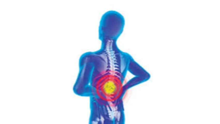
Do your legs feel heavy, weak or painful after walking just a few hundred metres, and do you find relief only when you sit down or lean forward on a shopping trolley? This typical pattern, called neurogenic claudication, is the hallmark of lumbar spinal stenosis - one of the most common reasons adults over fifty consult a spine surgeon. Stenosis develops slowly, often over years, as the spinal canal gradually narrows and squeezes the nerves inside it. Left untreated, it can shrink a person's world, limiting walking, travel, work and independence.
The good news is that spinal stenosis is very treatable. Many patients improve with physiotherapy and medicines, and for those who need surgery, modern endoscopic and minimally invasive decompression can free the nerves through a small incision, with far less muscle damage than traditional laminectomy. Dr. (Prof.) Sumiet Snha, AIIMS-trained neurosurgeon and Principal Director - Neurosurgery & Spine Surgery at Max Hospital, Dwarka, New Delhi, has extensive experience in treating single-level and multi-level stenosis in the neck and lower back, including in elderly patients with other medical conditions.
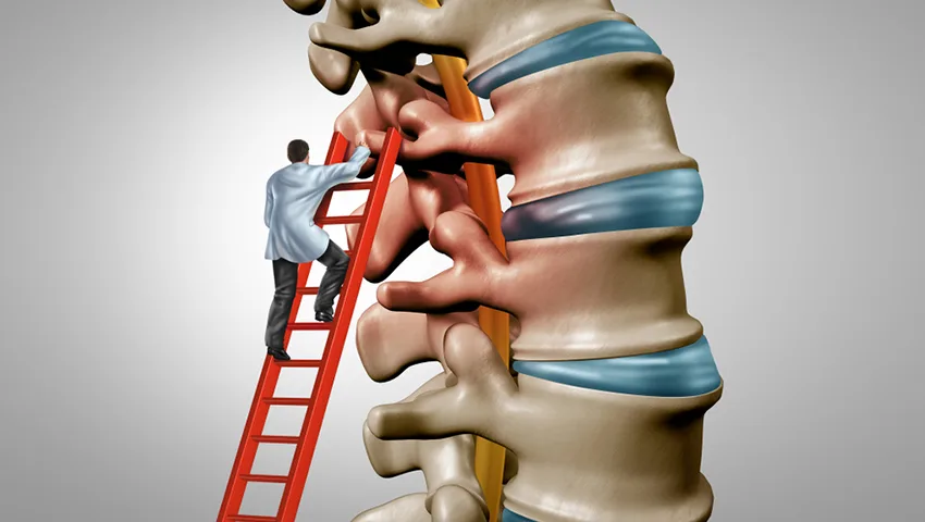
What Is Spinal Stenosis?
"Stenosis" means narrowing. Spinal stenosis is narrowing of the spaces within the spine through which the spinal cord and nerve roots travel. It can affect three main areas:
Central canal stenosis
Narrowing of the main canal in the middle of the spine, compressing the spinal cord (neck) or the bundle of nerve roots (lower back).
Lateral recess stenosis
Narrowing of the gutter at the side of the canal where the nerve root begins its exit, often caused by facet joint overgrowth.
Foraminal stenosis
Narrowing of the foramen - the opening through which each nerve root leaves the spine - typically from disc height loss and bone spurs.
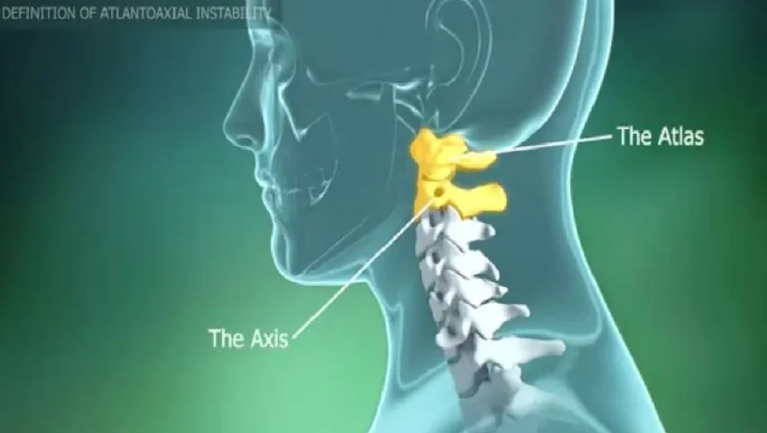
Cervical stenosis
Narrowing in the neck that may compress the spinal cord (myelopathy) or nerve roots (radiculopathy), affecting the arms, hands and walking.
Lumbar (lower back) stenosis is most common, followed by cervical (neck) stenosis. Thoracic stenosis is less frequent. The degree of narrowing seen on the MRI does not always match the severity of symptoms, which is why clinical assessment is as important as imaging.
Why Does the Spinal Canal Become Narrow?
Age-related degeneration
The most common cause is the natural wear and tear of the spine. Over the years, discs lose water and height and bulge backwards. The ligamentum flavum - a ligament lining the back of the canal - thickens and buckles inwards. The facet joints enlarge with arthritis and form bone spurs (osteophytes). Each change alone may be small, but together they can significantly reduce the space available for the nerves.
Degenerative spondylolisthesis
In some people, one vertebra slips forward over the one below, most often at L4-L5. This slip further narrows the canal and can cause both stenosis and instability.
Congenital narrow canal
Some people are born with a relatively narrow canal. They may develop symptoms earlier in life, even with modest degenerative changes.
Other causes
- Disc herniation superimposed on an already narrow canal.
- Previous spine surgery or injury with scar tissue or instability.
- Ossification of the posterior longitudinal ligament (OPLL), especially in the neck.
- Paget's disease, tumours or infection - uncommon causes that must be excluded.
Symptoms of Lumbar Spinal Stenosis
- Pain, heaviness, cramping or tiredness in the buttocks and legs when standing or walking.
- Relief on sitting, bending forward or lying down - bending forward opens up the canal.
- Reduced walking distance that gradually decreases over months.
- Numbness, tingling or a sensation of "walking on cotton".
- Weakness in the legs or foot drop in advanced cases.
- Lower back pain, often less prominent than the leg symptoms.
Many patients notice that they can cycle or walk uphill more comfortably than downhill, because the forward-leaning posture enlarges the canal. This helps doctors distinguish nerve-related (neurogenic) claudication from vascular claudication caused by poor blood supply to the legs, which improves simply by standing still.
Symptoms of Cervical Spinal Stenosis
- Neck pain and stiffness.
- Pain, numbness or tingling radiating into the shoulders, arms and hands.
- Clumsiness of the hands - difficulty buttoning clothes, writing or holding small objects.
- Unsteady walking, loss of balance or a feeling of stiffness in the legs.
- In severe cases, bladder or bowel disturbance.
When stenosis becomes urgent
Cervical stenosis that compresses the spinal cord (cervical myelopathy) tends to progress in a step-wise manner and can cause permanent disability if neglected. Rapidly worsening weakness, falls, loss of hand function or bladder problems need prompt specialist assessment. In the lower back, sudden loss of bladder or bowel control or numbness in the saddle region is an emergency.
Cervical Myelopathy - Why Neck Stenosis Needs Special Attention
When stenosis in the neck compresses the spinal cord itself, the condition is called cervical spondylotic myelopathy. It is the most common cause of spinal cord dysfunction in adults over fifty, yet it is frequently missed because its early signs are subtle and are often attributed to ageing, arthritis or diabetes. Patients may notice that their handwriting has changed, that they fumble with coins or buttons, that their hands feel numb and "not their own", or that they need to hold the railing when climbing stairs. Family members sometimes notice a wide-based or unsteady walk before the patient does.
Unlike lumbar stenosis, which affects nerve roots and often fluctuates, myelopathy reflects injury to the spinal cord, and damaged cord tissue has limited ability to recover. For this reason, moderate to severe or progressive myelopathy is usually treated surgically, with the aim of halting deterioration and allowing whatever recovery is possible. Mild myelopathy may be observed carefully with regular examinations, but patients are advised to avoid activities with a high risk of neck injury, since even a minor fall can cause a sudden worsening in a narrowed canal. The choice between an anterior operation (from the front of the neck), a posterior operation (from the back) or a combined approach depends on the number of levels involved, the alignment of the neck and where the compression lies.
How Spinal Stenosis Is Diagnosed
- Clinical history - the walking pattern, posture-related relief and the time course of symptoms.
- Physical and neurological examination - strength, reflexes, sensation, balance and gait; checking foot pulses to rule out vascular problems.
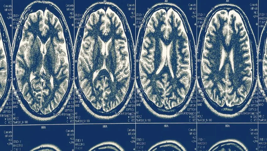
- MRI scan - the investigation of choice, showing the degree and level of narrowing and the condition of discs and ligaments.
- Standing and dynamic X-rays - to identify spondylolisthesis and instability, which influence the surgical plan.
- CT scan or CT myelogram - to assess bone spurs, calcification, OPLL or when MRI is not possible (for example, with some pacemakers).
- Nerve conduction studies - where peripheral neuropathy (common in diabetics) needs to be distinguished from nerve root compression.
- Arterial Doppler - if vascular claudication is suspected.
Non-Surgical Treatment of Spinal Stenosis
For mild to moderate symptoms, conservative treatment is always the first step and helps a large proportion of patients live comfortably.
Physiotherapy
Flexion-based exercises, core strengthening, stretching, balance training and stationary cycling to maintain fitness without aggravating symptoms.
Medicines
Pain relievers and nerve-pain medicines, used carefully in older adults with attention to kidney function and other conditions.
Epidural steroid injections
Image-guided injections can reduce nerve inflammation and provide relief for weeks to months in selected patients.
Lifestyle measures
Weight reduction, use of a walking stick or trolley, planned rest breaks and avoiding long periods of standing.
Stenosis does not usually "cure" itself because the narrowing is structural, but symptoms can remain stable for long periods. Surgery is considered when walking distance and quality of life become unacceptably limited despite adequate conservative care, or when there is progressive neurological deficit.
Surgical Options for Spinal Stenosis
The goal of surgery is decompression - to create more space for the nerves by removing the thickened ligament, overgrown bone and bulging disc material that are pressing on them. When the spine is also unstable, stabilisation (fusion) may be added.
1. Endoscopic decompression (unilateral approach, bilateral decompression)
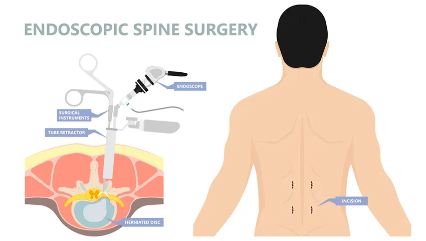
Through a small incision on one side, the endoscope is directed towards the canal. The surgeon removes the thickened ligamentum flavum and trims the overgrown facet joint, then angles the endoscope across the midline to decompress the opposite side as well - the so-called "over-the-top" technique. Muscles and the midline ligaments that give stability are preserved. This approach is especially valuable for elderly patients and those with medical conditions who may not tolerate a larger operation.
2. Microscopic / tubular decompression
A small tubular retractor and the operating microscope are used to achieve the same decompression. It is a well-established minimally invasive alternative.
3. Laminectomy
The traditional open operation, removing the lamina (the roof of the canal). It may still be appropriate for extensive multi-level stenosis or specific anatomical situations.
4. Decompression with minimally invasive fusion (MIS-TLIF)
If there is significant spondylolisthesis or instability, screws and an interbody cage are placed through small incisions to stabilise the segment while decompressing the nerves.
5. Cervical procedures
For neck stenosis, options include anterior cervical discectomy and fusion (ACDF), cervical disc replacement in selected patients, corpectomy, laminoplasty or posterior decompression and fixation, depending on the number of levels, the alignment of the neck and the location of compression.
The Endoscopic Decompression Procedure
- Assessment and planning - MRI and X-rays are studied to decide the levels, side of approach and whether fusion is needed.
- Anaesthesia and positioning - usually general anaesthesia; the patient lies face-down on a special frame that keeps the abdomen free.
- X-ray localisation - the correct level is confirmed with fluoroscopy.
- Portal placement - a small incision is made, muscles are dilated and the working channel is docked on the lamina.
- Decompression under HD vision - bone is drilled precisely, the thickened ligament is removed and the nerve roots on both sides are freed.
- Final check - the dura (nerve covering) and nerve roots are inspected to confirm adequate space.
- Closure - a few stitches; often no drain is needed.
Patients are usually encouraged to walk the same day or the day after surgery. Hospital stay is typically short, depending on the number of levels and the patient's general health.
Advantages of Endoscopic Stenosis Surgery
- Small incision and minimal muscle damage.
- Preservation of the midline structures that support spinal stability, reducing the chance of post-operative instability.
- Less blood loss and lower post-operative pain.
- Early walking and shorter hospital stay.
- Particularly suitable for elderly patients and those with diabetes, heart or lung disease.
- Faster return to independent daily activities.
Risks and Realistic Expectations
Decompression surgery is generally very effective in relieving leg pain and improving walking distance. Back pain from arthritis may improve less predictably. Possible risks include dural tear, infection, bleeding, temporary numbness or tingling, incomplete relief and, over time, the development of stenosis at other levels. Nerves that have been compressed for many years may not recover completely, which is one reason why timely treatment matters. Your surgeon will discuss your individual risk profile in detail.
Recovery After Spinal Stenosis Surgery
General recovery guide
- Day 0-1: walking with assistance; many patients notice that leg heaviness has eased.
- Day 1-3: discharge home with instructions on wound care and activity.
- Week 2: wound review; increasing walking distance daily.
- Week 3-8: physiotherapy focusing on core strength, posture and endurance.
- Month 3 onwards: most patients have resumed normal daily activities; fusion patients follow a longer timeline.
Walking is the best exercise after decompression. Gradually increasing your distance each day builds stamina and confidence. Avoid heavy lifting and repeated bending until your surgeon clears you.
Spinal Stenosis in the Elderly - Special Considerations
Many patients with stenosis are in their sixties, seventies or even eighties. Families often worry whether surgery is safe at this age. With careful pre-operative evaluation, optimisation of heart, lung, kidney and blood sugar status, and the use of minimally invasive techniques, surgery can be performed safely in carefully selected older patients. Restoring the ability to walk independently has a major positive effect on overall health - improving heart fitness, blood sugar control, mood and social life, and reducing the risk of falls. The decision is always individual and is taken jointly with the patient and family.
Why Consult Dr. (Prof.) Sumiet Snha?
- MBBS, MS, DNB, MCh (AIIMS, New Delhi), FACS - with over 25 years of experience in spine and brain surgery.
- Former Professor of Neurosurgery, AIIMS, New Delhi.
- Certified teaching faculty for AO Spine International and trained with renowned spine surgeons abroad.
- Expertise in the full spectrum of stenosis treatment - endoscopic, tubular, open and fusion surgery - so the choice of technique is based on your needs, not on the limits of a single method.
- A conservative-first philosophy: surgery is advised only when it is likely to improve quality of life.
- State-of-the-art facilities at Max Super Speciality Hospital, Dwarka, New Delhi.
Cost of Spinal Stenosis Surgery in Delhi - What Affects It?
- Number of levels requiring decompression.
- Whether fusion with screws and cages is needed.
- Endoscopic vs microscopic vs open technique.
- Length of hospital stay and room category.
- Associated medical conditions requiring additional care.
- Insurance and cashless eligibility.
A clear, itemised estimate is provided after the consultation and imaging review.
Exercises and Habits That Help People With Stenosis
- Knee-to-chest stretches and pelvic tilts, which open the canal and relieve pressure.
- Stationary cycling - a leaning-forward posture allows longer exercise without symptoms.
- Aquatic exercise - buoyancy reduces load on the spine.
- Core strengthening to support the lower back.
- Weight control to reduce mechanical stress on the spine.
- Avoiding prolonged standing and excessive arching of the back.
Always learn exercises from a trained physiotherapist first, especially if you have balance problems or osteoporosis.
Understanding Your MRI Report
MRI reports for stenosis often contain terms that can sound alarming. "Disc desiccation" simply means the disc has lost some water with age. "Diffuse disc bulge" describes a broad-based bulging of the disc margin, which is very common. "Ligamentum flavum hypertrophy" means the ligament at the back of the canal has thickened. "Facet arthropathy" refers to arthritis of the small joints of the spine. "Thecal sac indentation" and "canal compromise" describe how much the nerve sac is being narrowed. Words like "mild", "moderate" and "severe" grade the narrowing, but the grade alone does not decide treatment - your symptoms and examination do. Dr. Snha explains your scan in plain language so that you understand exactly what it shows and what it does not.
Conditions That Can Mimic Spinal Stenosis
Leg pain on walking is not always caused by the spine. An experienced spine specialist carefully rules out other conditions, because treating the wrong cause leads to disappointment. The common look-alikes include:
Peripheral arterial disease
Narrowed leg arteries cause calf cramping on walking that settles quickly on standing still, without needing to sit or bend. Foot pulses may be weak; a Doppler study confirms it.
Hip arthritis
Pain in the groin or thigh, stiffness while putting on socks and pain on rotating the hip. Hip X-rays and examination help separate it from spinal pain.
Diabetic neuropathy
Burning, numbness and tingling in both feet in a "stocking" pattern, present even at rest. Nerve conduction tests are helpful.
Sacroiliac joint or piriformis problems
Buttock pain that may radiate to the thigh, often provoked by specific movements and tender on examination.
It is also common for two problems to coexist - for example, stenosis together with knee or hip arthritis. In such cases the treatment is planned in a sequence that gives the greatest improvement in walking and comfort.
Preparing for Spinal Stenosis Surgery
Good preparation makes surgery safer and recovery smoother. Before your operation, the team will guide you through the following:
- Medical fitness check - blood tests, ECG, chest X-ray and, where needed, echocardiography or a physician's review, particularly for patients with diabetes, heart disease or lung problems.
- Medicine review - blood thinners such as aspirin, clopidogrel or newer anticoagulants may need to be stopped or bridged under the supervision of your cardiologist or physician.
- Blood sugar control - well-controlled sugar reduces the risk of wound infection and helps healing.
- Stopping smoking - even a few weeks without smoking improves wound and bone healing.
- Home preparation - arrange a firm chair with arms, keep commonly used items at waist height and remove loose rugs to prevent falls.
- Understanding the plan - know your expected length of stay, the exercises you will start and who to call if you have questions after discharge.
Life After Decompression - Practical Guidance
Most patients are surprised by how quickly they can walk after minimally invasive decompression. Still, the tissues need time to heal. In the first two weeks, keep the wound clean and dry, walk several times a day and avoid lifting anything heavier than a light bag. Sitting is permitted, but change position every 30 to 45 minutes. Sleep in whichever position is comfortable; a pillow between the knees helps when lying on the side.
From the third week, a physiotherapist introduces gentle core activation, hamstring stretches and progressive walking or stationary cycling. By six to eight weeks, most people who had decompression alone return to routine household work, social activities and travel. Those who had fusion follow a slower plan while the bone heals. Some residual numbness in the feet is common if it had been present for a long time before surgery; it often improves gradually over months. Follow-up visits allow the surgeon to track recovery and adjust the exercise programme.
In the longer term, staying physically active is the best protection for your spine. Regular walking, maintaining a healthy weight, strengthening the core and avoiding smoking all slow further degeneration. If new symptoms appear years later, they can usually be managed with the same step-wise approach - conservative care first, and minimally invasive surgery only if needed.
Glossary of Common Terms
- Neurogenic claudication: leg pain, heaviness or weakness on walking caused by nerve compression in the spine.
- Laminotomy: removal of a small part of the lamina to create space for the nerves.
- Laminectomy: removal of the entire lamina over one or more levels.
- Foraminotomy: widening of the exit opening of a nerve root.
- Spondylolisthesis: forward slippage of one vertebra over another.
- Fusion: joining two vertebrae with bone graft, usually supported by screws and a cage, to stop abnormal movement.
- Myelopathy: dysfunction of the spinal cord due to compression, usually in the neck.
A Typical Patient Journey (Illustrative Example)
The following composite example illustrates a common course of treatment. Imagine a retired teacher in her late sixties who notices that her morning walks in the park are becoming shorter each month. After about ten minutes her legs feel heavy and numb, and she has to sit on a bench before continuing. Physiotherapy and medicines help for a while, but eventually she can barely manage a trip to the market. Her MRI shows severe narrowing at L4-L5 with no instability. After discussing the options, she undergoes endoscopic decompression through a small incision on one side, with both sides of the canal freed. She walks the same evening and goes home the next day. Over the following weeks, guided by her physiotherapist, she gradually increases her walking distance and returns to her morning routine. This pattern is common, though every patient's journey is individual, and the right plan always depends on the specific findings and goals.
Tandem Stenosis - When the Neck and Lower Back Are Both Narrowed
Some patients have significant narrowing of the spinal canal in both the neck (cervical) and lower back (lumbar) regions at the same time - a situation called tandem stenosis. It is more common than many people realise, particularly in older adults and those with a congenitally narrow canal. The difficulty is that the symptoms can overlap: leg weakness, imbalance and difficulty walking may come from compression of the spinal cord in the neck, from nerve compression in the lower back, or from both.
Clues that the neck is involved
- Clumsy hands, difficulty with buttons or handwriting.
- Unsteady, broad-based walking even when leg pain is mild.
- Brisk reflexes and other signs of spinal cord involvement on examination.
- Symptoms that do not ease on sitting or bending forward, unlike typical lumbar claudication.
Careful examination, followed by MRI of both regions, is the key to sorting out which area is responsible. When cervical cord compression is significant, it is generally addressed first, because an untreated compressed cord is vulnerable during positioning for lumbar surgery and carries a greater risk of lasting disability. The lumbar stenosis can then be treated, often endoscopically, once recovery from the neck operation allows. In selected patients the two procedures may be combined. Dr. Snha explains the recommended order and the reasons for it, so that patients understand why treating one region first may give the best overall result.
Frequently Asked Questions
Can spinal stenosis be treated without surgery?
Yes. Many patients with mild to moderate symptoms do well with physiotherapy, medicines, lifestyle changes and, in selected cases, epidural injections. Surgery is considered when symptoms significantly limit daily life or there is progressive weakness.
Is endoscopic surgery suitable for multi-level stenosis?
Endoscopic decompression can be performed at more than one level in selected cases. When many levels are involved or there is deformity or instability, other minimally invasive or open techniques may be more appropriate.
Will I need screws and rods?
Not always. Fusion is added only when there is significant instability, such as a mobile spondylolisthesis. Many patients need decompression alone.
How quickly will my walking improve?
Many patients notice an improvement in leg heaviness and walking distance within days to weeks. Numbness and weakness may take longer to recover.
Is surgery safe for people over 70?
With careful medical evaluation and minimally invasive techniques, decompression surgery can be performed safely in many older patients. The decision is individualised after assessing overall health.
Can stenosis come back after surgery?
The decompressed level usually stays open, but degenerative changes can develop at other levels over time. Staying active and maintaining a healthy weight helps protect the spine.
What is the difference between stenosis and a slip disc?
A slip disc is a localised herniation of disc material, often in younger adults, causing sudden sciatica. Stenosis is a gradual narrowing from multiple degenerative changes, usually in older adults, causing leg symptoms on walking.
Get Back On Your Feet
If walking has become difficult because of leg pain or heaviness, you do not have to accept it as a normal part of ageing. A careful evaluation can identify whether spinal stenosis is the cause and which treatment is right for you. Book an appointment with Dr. (Prof.) Sumiet Snha at Max Hospital, Dwarka, or learn more about endoscopic spine surgery and complex cervical spine conditions.