Our peripheral nerves are the body's wiring system - carrying commands from the brain and spinal cord to the muscles, and bringing back information about touch, temperature, position and pain. When a peripheral nerve is injured, the effects can be profound: a hand that cannot grip, a wrist that drops, a foot that drags, numbness that leads to unnoticed burns and cuts, or pain that never seems to stop. Peripheral nerve injuries are common - from glass cuts and machinery injuries to fractures, road accidents, injections, and nerve compression syndromes - yet they are often underestimated or treated too late.
Unlike the brain and spinal cord, peripheral nerves can regenerate, and with the right treatment at the right time, many patients regain useful or even near-normal function. Dr. (Prof.) Sumiet Snha, Founder Secretary of the Indian Society of Peripheral Nerve Surgery and a former Professor of Neurosurgery at AIIMS, New Delhi, has treated more than 250 complex nerve injuries. At Max Hospital, Dwarka, he provides comprehensive peripheral nerve care - from diagnosis and nerve studies to microsurgical repair, grafting, nerve transfers and rehabilitation.

Understanding Peripheral Nerves
A peripheral nerve is like a cable containing thousands of tiny wires (axons), each insulated by a myelin sheath and grouped into bundles called fascicles, all wrapped in protective layers of connective tissue. Some fascicles carry motor signals to muscles, others carry sensory information back to the spinal cord. Major peripheral nerves include the median, ulnar and radial nerves in the arm, the sciatic, peroneal (fibular) and tibial nerves in the leg, the femoral nerve in the thigh, and the facial nerve in the face. Injury to each produces a characteristic pattern of weakness and numbness that helps the specialist identify the nerve and the level of injury.
Common Causes of Peripheral Nerve Injury
Lacerations
Cuts from glass, knives, sheet metal or machinery, especially at the wrist, forearm and elbow.
Fractures and dislocations
E.g. radial nerve injury with fractures of the upper arm, peroneal nerve injury with knee dislocations.
Stretch and crush injuries
From road accidents, falls and industrial injuries.
Injection injuries
Improperly placed intramuscular injections in the buttock can injure the sciatic nerve - a preventable cause seen in India.
Iatrogenic injuries
Nerves may occasionally be injured during surgery, such as lymph node biopsy in the neck (spinal accessory nerve) or hip and knee operations.
Compression (entrapment)
Chronic pressure at narrow passages - carpal tunnel, cubital tunnel, fibular head - or from prolonged pressure during unconsciousness.
Gunshot and blast injuries
Often a combination of stretch, crush and thermal damage.
Tumors
Nerve sheath tumors and other masses pressing on nerves - see our nerve tumor page.
Common Nerve Injuries and Their Symptoms
Radial nerve injury - wrist drop
The radial nerve controls straightening of the elbow, wrist and fingers. Injury, commonly with a fracture of the humerus (upper arm bone) or from pressure (so-called "Saturday night palsy"), causes wrist drop - inability to lift the wrist and fingers - with numbness on the back of the hand.
Median nerve injury
The median nerve supplies the muscles that bend the thumb, index and middle fingers and oppose the thumb, and sensation to the thumb side of the palm. Injury at the wrist - often from glass cuts - causes numbness of the thumb, index and middle fingers and weakness of thumb movements. Chronic compression at the wrist causes carpal tunnel syndrome.
Ulnar nerve injury - claw hand
The ulnar nerve controls many of the small muscles of the hand, essential for fine movements and grip strength. Injury causes numbness of the little and ring fingers, weakness of grip, wasting of the hand muscles, and in time a "claw hand" deformity. Compression at the elbow (cubital tunnel syndrome) is common.
Peroneal (fibular) nerve injury - foot drop
The common peroneal nerve winds around the head of the fibula just below the knee, where it is vulnerable to injury from knee trauma, fractures, tight plasters, prolonged leg crossing, squatting and rapid weight loss. Injury causes foot drop - inability to lift the foot and toes, leading to tripping and a high-stepping gait - with numbness on the top of the foot.
Sciatic nerve injury
Injury to the large sciatic nerve - from hip fractures, hip surgery, gunshot wounds or misplaced injections - can cause weakness below the knee, foot drop, and numbness and pain in the leg and foot.
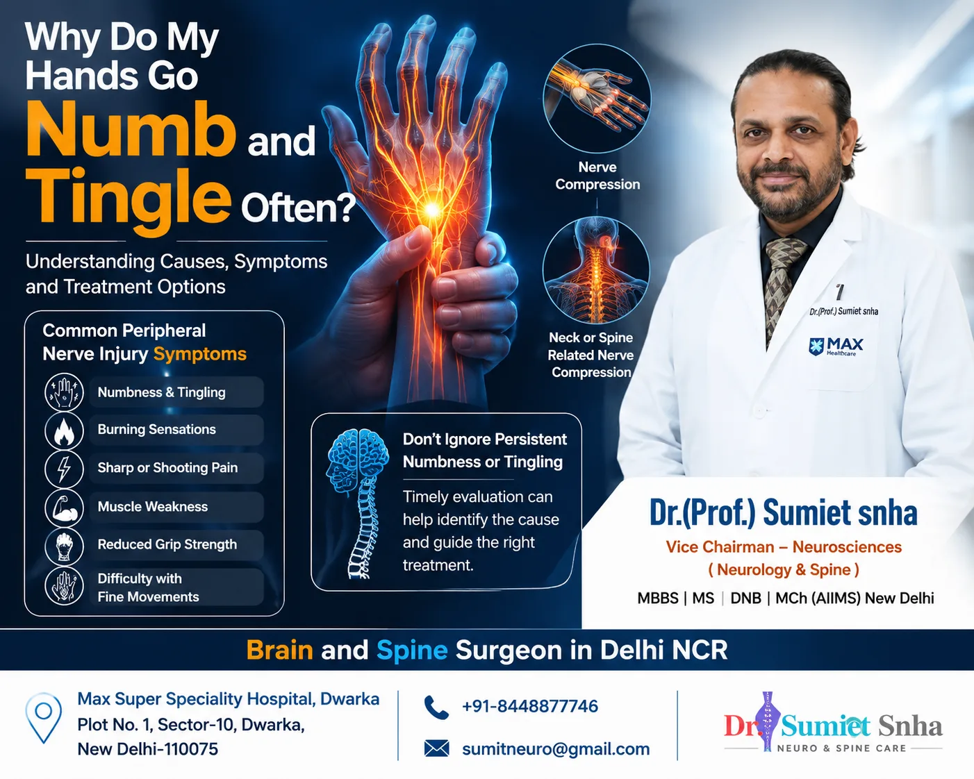
Symptoms That Suggest a Nerve Injury
- Weakness or paralysis of specific muscles - difficulty lifting the wrist or foot, gripping, pinching or spreading the fingers.
- Numbness, tingling or "pins and needles" in a defined area of skin.
- Muscle wasting (thinning) over weeks to months.
- Burning, shooting or electric-shock pain, sometimes triggered by light touch.
- A tender lump along a nerve after injury (neuroma) that causes electric shocks when tapped.
- Loss of sweating in the affected area.
- Repeated injuries or burns to numb areas.
After a cut, check the fingers
If a cut on the wrist, forearm, palm or fingers is followed by numbness of any finger or difficulty moving the fingers or thumb, a nerve or tendon may be cut. These injuries should be repaired early by a specialist - ideally within days. Simply stitching the skin can leave a severed nerve unrepaired, and delayed repair gives poorer results.
How Nerve Injuries Are Diagnosed
- Clinical examination - careful testing of each muscle and area of sensation identifies which nerve is injured and at what level. The Tinel sign (tingling produced by tapping along the nerve) can help locate the injury and track regeneration.
- Nerve conduction studies and EMG - measure how well nerves conduct signals and whether muscles have lost their nerve supply; typically done 3-4 weeks after injury and repeated to assess recovery.
- High-resolution ultrasound - shows the nerve's continuity, swelling, neuromas and compression; useful in the clinic and during follow-up.
- MR neurography - specialised MRI that visualises nerves and muscles.
- X-rays and CT - for associated fractures.
Treatment of Peripheral Nerve Injuries
The choice of treatment depends on the type of injury, its mechanism and the time since injury.
Observation and therapy
Injuries where the nerve is in continuity - such as stretch or pressure injuries - often recover on their own. They are monitored clinically and with nerve studies. Splints (for example, a wrist splint for wrist drop or an ankle-foot orthosis for foot drop) maintain function and prevent deformity while waiting, and physiotherapy keeps the joints supple. If there is no sign of recovery within the expected time (usually 3-4 months), surgical exploration is considered.
Primary nerve repair
Clean-cut nerves are best repaired directly, end-to-end, under the operating microscope, ideally within the first few days. The nerve ends are trimmed and aligned so that corresponding fascicles meet, and they are joined with sutures finer than a hair, avoiding tension.
Nerve grafting
When there is a gap between nerve ends - because of tissue loss or retraction in delayed repair - a segment of a less important sensory nerve (usually the sural nerve from the back of the leg) is used to bridge it. In some small gaps, nerve conduits or processed nerve allografts may be options.
Neurolysis
When a nerve is intact but trapped in scar tissue or compressed, it is freed (neurolysis). Intra-operative nerve action potential recordings help decide whether a segment is regenerating and can be left alone, or needs to be removed and grafted.
Nerve transfers
A healthy nerve branch with a less critical function is connected to the paralysed nerve close to its target muscle. For example, in high ulnar nerve injuries, a branch of the anterior interosseous nerve can be transferred to the motor branch of the ulnar nerve in the hand; in radial nerve palsy, branches of the median nerve can restore wrist and finger extension; and in foot drop, a branch of the tibial nerve can be transferred to the deep peroneal nerve in selected cases. Nerve transfers shorten the regeneration distance and are particularly useful for high injuries and late presentations.
Tendon transfers
When nerve recovery is not possible or has been incomplete, working muscles can be redirected to restore key functions - for example, tendon transfers for wrist drop or foot drop can provide excellent functional results.
Nerve Compression (Entrapment) Syndromes
Not all nerve injuries are sudden. Many develop gradually when a nerve is compressed at a narrow anatomical passage. These entrapment neuropathies are very common and highly treatable.
Carpal tunnel syndrome
Compression of the median nerve at the wrist causing numbness and tingling in the thumb, index and middle fingers, worse at night, and later weakness. Treated with splints and injections, or a simple carpal tunnel release operation.
Cubital tunnel syndrome
Compression of the ulnar nerve at the elbow causing numbness of the ring and little fingers and hand weakness. Treated by avoiding elbow flexion pressure or by decompression/transposition surgery.
Peroneal nerve entrapment
Compression at the fibular head causing foot drop, often after weight loss, leg crossing or squatting. Decompression can restore function.
Thoracic outlet syndrome
Compression of the brachial plexus near the neck and first rib, causing arm pain, numbness and weakness.
Tarsal tunnel syndrome
Compression of the tibial nerve at the ankle causing burning pain in the sole.
Meralgia paraesthetica
Compression of a sensory nerve in the thigh causing burning numbness on the outer thigh.
Early diagnosis with nerve conduction studies and ultrasound, and timely decompression when needed, prevent permanent muscle wasting and loss of sensation.
Painful Neuromas and Nerve Pain
When a nerve is cut and not repaired, or after some injuries and amputations, the regenerating fibres can form a tangled, sensitive bundle called a neuroma. Neuromas cause sharp, electric pain when touched or pressed and can make it difficult to wear shoes, use a prosthesis or hold objects. Treatment options include medicines, targeted injections, and surgical procedures such as neuroma excision with nerve repair, burying the nerve end in muscle, or newer techniques like targeted muscle reinnervation that give the nerve "something to do". Chronic nerve pain after injury may also benefit from neuromodulation in selected cases - see our spinal cord stimulator page.
What Determines the Outcome of Nerve Surgery?
- Timing - earlier repair generally gives better results; muscles lose their capacity to accept new nerve supply after about 12-18 months.
- Age - children recover better than adults.
- Level of injury - injuries closer to the target muscles recover better than those far away.
- Type of nerve - purely motor or purely sensory nerves recover better than mixed nerves.
- Mechanism - clean cuts do better than crush, stretch or blast injuries.
- Gap length - short gaps and tension-free repairs yield better results.
- Rehabilitation - committed physiotherapy and sensory re-education significantly improve outcomes.
Recovery and Rehabilitation After Nerve Surgery
General timeline
- First 3-4 weeks: the repaired limb is protected in a splint to avoid tension on the repair.
- Weeks 4-12: gradual mobilisation of joints; splints to maintain function (e.g. wrist or foot splints).
- Months 3-12: nerve regeneration progresses at roughly a millimetre per day; the Tinel sign advances along the nerve.
- Recovery of muscle function: first flickers of movement may appear within months, with continued improvement over 1-2 years.
- Sensory re-education: training the brain to interpret new sensory signals improves hand function.
Throughout recovery, protect numb areas from burns and injuries, keep joints mobile and attend regular follow-up to monitor progress and plan any secondary procedures.
Nerve Injuries From Injections - A Preventable Problem
In many parts of India, intramuscular injections in the buttock are commonly given for fever, pain or weakness. If an injection is placed too low or too close to the midline, it can injure the sciatic nerve, causing immediate burning pain radiating down the leg, followed by weakness and foot drop. Children and thin adults are particularly vulnerable. Treatment depends on the severity: many injuries improve with time and therapy, while others need exploration and neurolysis or reconstruction. Prevention is simple - injections should be given only when truly necessary, preferably by trained staff, and in the upper outer quadrant of the buttock or the thigh, well away from the course of the sciatic nerve.
Facial Nerve Injuries
The facial nerve controls the muscles of expression. It can be injured by facial lacerations, skull base fractures, surgery for acoustic neuroma or parotid tumors, and Bell's palsy (a viral inflammatory condition that usually recovers without surgery). When the nerve is cut by trauma, early microsurgical repair gives the best results. When the proximal nerve is not available, nerve transfers - such as from the nerve to the masseter muscle or the hypoglossal nerve - can restore facial tone and a smile. Eye protection is essential while recovery takes place.
Spinal Accessory Nerve Injury
This nerve controls the trapezius muscle, which helps lift and stabilise the shoulder. It runs superficially in the neck and can be injured during lymph node biopsies or neck surgery, causing shoulder pain, drooping, and difficulty raising the arm. Early recognition and repair or grafting can restore function; tendon transfers are an option for late cases.
Why Choose Dr. (Prof.) Sumiet Snha for Nerve Injuries?
- Founder Secretary, Indian Society of Peripheral Nerve Surgery - a leader in the field in India.
- More than 250 complicated peripheral nerve and brachial plexus operations with high success rates.
- Former Professor of Neurosurgery at AIIMS, New Delhi, and the JPNA Apex Trauma Centre.
- Expertise in microsurgical repair, grafting, nerve transfers, neuroma surgery and entrapment decompression.
- Certified AO Spine and ATLS faculty with deep trauma experience.
- Integrated physiotherapy, occupational therapy and electrodiagnostic services at Max Super Speciality Hospital, Dwarka.
Factors That Affect the Cost of Nerve Surgery
- Type of procedure - decompression, primary repair, grafting, nerve transfer or tendon transfer.
- Number of nerves involved and duration of surgery.
- Investigations such as nerve conduction studies, ultrasound and MRI.
- Hospital stay (often short) and rehabilitation needs.
- Insurance or accident claim coverage.
Radial Nerve Palsy With Humerus Fractures - A Closer Look
The radial nerve spirals around the back of the humerus in a groove, lying directly against the bone. Fractures of the shaft of the humerus, particularly in the middle and lower thirds, can stretch, bruise or occasionally cut the nerve. Many such injuries are neurapraxias or axonotmesis that recover spontaneously over three to six months, so if the fracture is treated without surgery and the nerve palsy was present from the time of injury, doctors usually monitor it with a wrist splint and physiotherapy. However, if the fracture is open, if the palsy develops after manipulation or fixation, or if there are no signs of recovery on clinical examination and nerve studies within the expected time, surgical exploration is indicated. Findings range from a nerve trapped in the fracture or scar, which can be freed, to a nerve that is torn and needs grafting. When recovery remains incomplete, well-established tendon transfers reliably restore wrist and finger extension. Early referral to a nerve specialist ensures that the window for nerve surgery is not missed.
Diabetes and Peripheral Nerves
Diabetes affects peripheral nerves in several ways. Diabetic polyneuropathy causes numbness, tingling and burning in the feet and hands in a symmetrical "stocking and glove" pattern and increases the risk of foot ulcers. People with diabetes are also more susceptible to entrapment neuropathies such as carpal tunnel syndrome, cubital tunnel syndrome and peroneal nerve compression, because their nerves are more vulnerable to pressure. Distinguishing a treatable compression from general diabetic neuropathy is important, since decompression can relieve symptoms caused by entrapment. Good blood sugar control, daily foot inspection, proper footwear and regular check-ups are essential for protecting nerves in people with diabetes.
Sensory Re-education and Hand Therapy
After nerve repair in the hand, regenerating fibres do not always reconnect with exactly the same skin areas they supplied before the injury. The brain initially receives confusing signals, so touch may feel strange, misplaced or unpleasant. Sensory re-education is a structured programme that retrains the brain to interpret these new signals. It begins with recognising where touch is applied while watching, then without looking, and progresses to identifying textures, shapes and everyday objects. Desensitisation techniques help reduce hypersensitivity. Combined with exercises for strength and dexterity guided by a hand therapist, sensory re-education significantly improves functional hand use after nerve injuries.
Nerve Injuries in Children
Children's nerves generally regenerate faster and more completely than those of adults, and a child's brain is better able to adapt to altered nerve connections. As a result, the outcome after nerve repair in children is often better than in adults with a similar injury. However, nerve injuries in children are easily missed. A young child may not be able to describe numbness, and may simply avoid using the hand or limp slightly. After a glass cut to the wrist or a fracture around the elbow - a common injury in children who fall from swings or trees - parents and doctors should specifically check finger movement and sensation.
A helpful clue in young children is the wrinkle test: skin supplied by a healthy nerve normally wrinkles after soaking in warm water for several minutes, while skin supplied by a cut nerve often does not. Watching how the child picks up small objects can also reveal weakness. Fractures around the elbow, particularly supracondylar fractures, can stretch or bruise the median, radial or ulnar nerves; most such injuries recover spontaneously, but recovery must be monitored and exploration considered if it does not progress as expected. Birth-related brachial plexus injuries are a separate topic covered on our brachial plexus page and paediatric neurosurgery page.
Returning to Work, Driving and Sport
One of the first questions patients ask is when they can return to normal activities. The answer depends on the nerve involved, the type of surgery and the demands of the person's job or sport. After a simple decompression, such as carpal tunnel release, many people return to desk work within days to a couple of weeks. After nerve repair or grafting, the repaired nerve must be protected from tension for several weeks, and the limb is often kept in a splint or brace during this period.
- Office work - often possible within a few weeks of repair, with adjustments such as voice software or a one-handed keyboard setup.
- Manual work - heavy lifting, gripping tools and vibrating equipment are usually avoided until healing and early recovery allow; a graded return is planned with the therapist.
- Driving - you should drive only when you can control the vehicle safely, including emergency braking and steering, and when splints no longer restrict movement. Foot drop affecting the pedal foot needs particular caution.
- Sport - non-contact exercise such as walking or stationary cycling can begin early; contact sports and heavy gym work are resumed gradually after discussion with the surgeon.
Protecting numb skin during recovery
While sensation is returning, numb areas cannot warn you of injury. Check the skin daily for cuts, blisters or redness, test bath water with an unaffected hand, use gloves when cooking or handling hot objects, and wear well-fitting footwear if the foot is affected. These simple habits prevent burns and wounds that can heal slowly.
Nerve, Spine or Muscle? Finding the True Source of Weakness
Weakness or numbness in a limb does not always come from an injured peripheral nerve. The same symptoms can arise from a trapped nerve root in the spine, a problem in the brain, or a disease of the muscles themselves - and the treatment for each is entirely different. A common example is foot drop: it may be caused by compression of the peroneal nerve at the knee, often after prolonged squatting, crossing the legs or weight loss, but it can equally result from an L5 nerve root pinched by a slipped disc in the lower back. Operating on the wrong site does not help the patient.
Careful examination is the first and most important tool. With a peroneal nerve problem, the muscle that turns the foot inwards is usually spared, whereas with an L5 root problem it is often weak as well, and back pain radiating down the leg may be present. Similarly, numbness in the little finger may come from the ulnar nerve at the elbow or from the C8 nerve root in the neck. Nerve conduction studies and EMG help locate the level of the problem, and MRI of the spine or nerve ultrasound is added where needed.
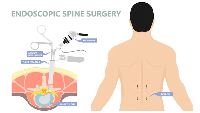
Because Dr. Snha treats both spinal and peripheral nerve conditions, both possibilities are considered together at the first consultation. Occasionally a patient has two problems at once - a so-called "double crush" - and both may need attention for the best recovery. For spinal causes, see our endoscopic spine surgery page.
Frequently Asked Questions
Can a damaged nerve heal on its own?
Nerves that are stretched or compressed but still intact often recover over weeks to months. Nerves that are cut or severely damaged need surgical repair to recover.
How soon should a cut nerve be repaired?
Ideally within the first few days. Delayed repair is still possible but results are generally better with early surgery.
How fast do nerves grow back?
Nerves regenerate at roughly one millimetre per day, or about an inch a month. Recovery time depends on the distance between the injury and the target muscles.
What is foot drop and can it be treated?
Foot drop is the inability to lift the front of the foot, often due to peroneal nerve injury or compression. Depending on the cause, treatment includes decompression, nerve repair, nerve transfer or tendon transfer.
Is carpal tunnel surgery a major operation?
No. Carpal tunnel release is a short procedure usually done under local anaesthesia, and most patients go home the same day.
Will I get full sensation back after nerve repair?
Sensory recovery varies. Many patients regain useful protective sensation; complete normal sensation is less common in adults but more likely in children.
What happens if nerve recovery is incomplete?
Secondary procedures such as tendon transfers can restore important functions even when nerve recovery is limited.
Restore Function - Seek Expert Care Early
If you have weakness, numbness or pain after an injury, a cut or a fracture - or symptoms of nerve compression - early specialist evaluation gives the best chance of recovery. Book a consultation with Dr. (Prof.) Sumiet Snha at Max Hospital, Dwarka, New Delhi. Related pages: brachial plexus injuries, nerve tumors and spasticity treatment.