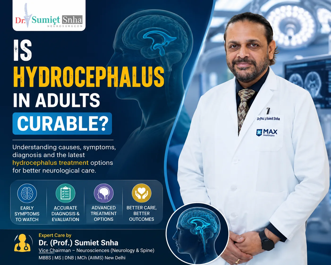
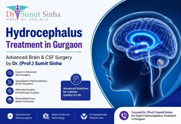
Hydrocephalus - often described in everyday language as "water in the brain" - is a condition in which cerebrospinal fluid (CSF) accumulates within the fluid-filled chambers of the brain, called ventricles. As the fluid builds up, the ventricles enlarge and press on the surrounding brain tissue. Hydrocephalus can affect newborn babies, children, adults and the elderly, and its causes and symptoms differ at different ages. Left untreated, it can cause developmental problems in children, loss of vision, memory and walking difficulties in adults, and, in its acute form, life-threatening pressure on the brain.
The good news is that hydrocephalus is very treatable. For decades, the standard treatment has been a shunt - a thin tube that drains excess fluid to another part of the body. In recent years, endoscopic third ventriculostomy (ETV) has become an important alternative for many patients, allowing the fluid to bypass the blockage through a tiny opening created inside the brain, often avoiding a lifelong implant. Dr. (Prof.) Sumiet Snha, a pioneer of minimally invasive and endoscopic neurosurgery and former Professor of Neurosurgery at AIIMS, New Delhi, offers both approaches at Max Hospital, Dwarka, selecting the right option for each patient.
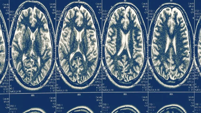
Understanding Cerebrospinal Fluid and the Ventricles
The brain is cushioned and nourished by cerebrospinal fluid, a clear, watery liquid produced continuously - roughly half a litre every day in adults - mainly by a structure called the choroid plexus within the ventricles. There are four ventricles: two lateral ventricles, one in each hemisphere, connected to a central third ventricle, which drains through a narrow channel called the aqueduct of Sylvius into the fourth ventricle at the back of the brain. From there, CSF flows out into the spaces around the brain and spinal cord and is finally absorbed into the bloodstream through structures called arachnoid granulations.
Because CSF is constantly being produced, any disruption of this circulation leads to accumulation. Hydrocephalus is broadly divided into two types based on where the problem lies:
Obstructive (non-communicating) hydrocephalus
Fluid flow is blocked within the ventricular system - most commonly at the aqueduct - by a narrowing, tumor, cyst or scar. The ventricles upstream of the block enlarge.
Communicating hydrocephalus
Fluid flows freely between the ventricles but is not absorbed properly, often after bleeding or infection, or in normal pressure hydrocephalus.
This distinction is central to treatment: ETV works best in obstructive hydrocephalus, while communicating hydrocephalus usually requires a shunt.
Causes of Hydrocephalus
In babies and children
- Aqueductal stenosis - congenital narrowing of the aqueduct.
- Spina bifida (myelomeningocele) with associated Chiari II malformation.
- Bleeding in the brain of premature babies (intraventricular haemorrhage).
- Infections - meningitis, including tubercular meningitis, which is an important cause in India.
- Brain tumors and cysts blocking fluid pathways, especially in the posterior fossa.
- Congenital malformations such as Dandy-Walker malformation.
In adults
- Brain tumors, colloid cysts and pineal region lesions.
- Subarachnoid haemorrhage from a ruptured aneurysm or head injury.
- Meningitis and other infections.
- Long-standing aqueductal stenosis presenting in adulthood (LOVA).
- Normal pressure hydrocephalus (NPH) in older adults.
Symptoms of Hydrocephalus
In infants
Because a baby's skull bones are not yet fused, the head can expand as fluid builds up. Parents and paediatricians may notice a rapidly increasing head size crossing the percentile lines on growth charts, a tense or bulging fontanelle (soft spot), widely separated skull sutures, prominent scalp veins, and the eyes deviating downwards ("sunsetting eyes"). The baby may be irritable, feed poorly, vomit, sleep excessively or have seizures, and developmental milestones may be delayed.
In older children and adults
Once the skull has fused, it cannot expand, so pressure rises more quickly. Typical symptoms are headaches that are often worse in the morning or on lying down, nausea and vomiting, blurred or double vision, swelling of the optic discs seen on eye examination (papilloedema), drowsiness, irritability, difficulty concentrating, a decline in school or work performance, balance problems and, in severe cases, reduced consciousness.
In older adults - Normal Pressure Hydrocephalus
Normal pressure hydrocephalus produces a classic triad of symptoms that develop gradually: difficulty walking (a slow, shuffling, wide-based gait, as if the feet are stuck to the floor), urinary urgency or incontinence, and memory and thinking problems. Because these symptoms are often attributed to ageing, Parkinson's disease or dementia, NPH is frequently missed - which is unfortunate, because it is one of the few treatable causes of dementia-like symptoms.
Signs of dangerously raised pressure
Severe headache with repeated vomiting, increasing drowsiness, confusion, double vision or seizures - particularly in someone with a known shunt or brain lesion - may indicate acutely raised intracranial pressure. This is an emergency requiring immediate medical attention.
How Hydrocephalus Is Diagnosed
- Clinical examination - including head circumference in infants, fontanelle assessment, eye examination for papilloedema, and neurological assessment.
- Cranial ultrasound - in infants with an open fontanelle, a quick, radiation-free way to assess ventricle size.
- CT scan - fast and widely available, showing ventricular enlargement and sometimes the cause.
- MRI - the most detailed test, showing the site of obstruction (such as aqueductal stenosis), tumors, cysts and CSF flow patterns; special sequences help decide whether ETV is suitable.
- For suspected NPH - a high-volume lumbar tap test or extended lumbar drainage: removing some CSF and assessing whether walking improves helps predict response to shunt surgery; gait videos and cognitive testing are done before and after.
- Additional tests - CSF analysis for infection, and ophthalmology assessment for vision.
Endoscopic Third Ventriculostomy (ETV) - Treatment Without a Shunt
ETV is a minimally invasive neurosurgical procedure that treats obstructive hydrocephalus by creating a new pathway for CSF to leave the ventricular system, bypassing the blockage. Here is how it works:
- Under general anaesthesia, a small incision is made on the scalp and a single burr hole (about a centimetre wide) is made in the skull, usually just behind the hairline.
- A thin neuroendoscope with a camera and light is passed gently through the brain into the enlarged lateral ventricle and then into the third ventricle.
- The thin floor of the third ventricle is identified between important landmarks.
- A small opening is carefully made in this floor and gently widened with a tiny balloon catheter.
- CSF can now flow directly from the third ventricle into the spaces around the brainstem, where it is absorbed normally - bypassing the aqueductal block.
- The endoscope is withdrawn, and the small incision is closed.
The procedure typically takes around an hour, and most patients go home within a few days. In the same sitting, the endoscope can also be used to obtain a biopsy of a tumor causing the blockage, remove a colloid cyst, or open (fenestrate) fluid-filled cysts - making it a versatile tool.
Advantages of ETV Over a Shunt
- No permanent implant - avoiding the lifelong risks of shunt blockage, infection and mechanical failure.
- More physiological - restores the natural flow and absorption of CSF.
- Lower risk of over-drainage problems.
- Single, small incision with a short hospital stay.
- Ability to address the cause - biopsy or removal of certain lesions in the same procedure.
- Freedom from "shunt anxiety" - families often live with constant worry about shunt malfunction.
Who Is a Good Candidate for ETV?
ETV is most successful in patients with obstructive hydrocephalus, particularly those caused by aqueductal stenosis or by tumors in the posterior fossa or pineal region. Success is higher in older children and adults than in young infants, whose absorption pathways may not yet be mature. Surgeons use scoring systems, such as the ETV Success Score, which consider age, cause and whether there was a previous shunt, to estimate the likelihood of success for each patient. In infants and in communicating hydrocephalus, ETV alone is less likely to succeed; in selected infants, ETV combined with choroid plexus cauterisation (reducing CSF production) may be considered. When the chance of success is low, a shunt is usually the better first choice.
Risks of ETV
ETV is generally safe in experienced hands, but possible risks include bleeding, infection, CSF leak, transient hormonal disturbances (such as altered sodium levels), temporary memory issues or eye movement problems, and, rarely, injury to important blood vessels at the floor of the third ventricle. Importantly, the new opening can close over time in a minority of patients, causing hydrocephalus to return - sometimes years later. For this reason, patients and families are taught to recognise symptoms of recurrence, and follow-up is continued long term. If ETV fails, it can sometimes be repeated, or a shunt can be placed.
Ventriculoperitoneal (VP) Shunt Surgery
A shunt remains the most widely used and reliable treatment for many forms of hydrocephalus, including communicating hydrocephalus, normal pressure hydrocephalus and hydrocephalus in young infants. A shunt system has three parts:
Ventricular catheter
A soft tube placed into the ventricle through a small hole in the skull.
Valve
Controls the pressure and amount of CSF drainage. Programmable valves can be adjusted from outside the body with a magnetic device, without further surgery.
Distal catheter
Tunnelled under the skin to the abdomen (peritoneal cavity), where the fluid is absorbed. Alternatives include the heart (VA shunt) or pleural cavity.
Lumboperitoneal shunt
In selected adults with communicating hydrocephalus, drainage from the lower spine to the abdomen.
Shunt surgery usually takes about an hour, and patients often go home within a few days. Shunts work well for many years in many patients, but because they are mechanical devices, they can malfunction or become infected, and some patients need revisions over their lifetime.
Signs of Shunt Malfunction
Everyone with a shunt - and their family - should know the warning signs of malfunction, which often resemble the original symptoms of hydrocephalus:
- Headache, nausea and vomiting.
- Drowsiness, irritability or confusion.
- In infants: bulging fontanelle, increasing head size, poor feeding, sunsetting eyes.
- Blurred or double vision.
- Decline in school performance, personality changes or new seizures.
- Fever, redness or swelling along the shunt track, or abdominal pain - possible infection.
Seek urgent medical attention if these symptoms appear. Early revision prevents serious complications.
Normal Pressure Hydrocephalus - A Treatable Cause of Walking and Memory Problems
Normal pressure hydrocephalus deserves special attention because it is common among older adults and is so often missed. In NPH, the ventricles enlarge even though CSF pressure measured by a lumbar puncture is normal or only intermittently raised. The enlarged ventricles stretch the nerve pathways that control walking, bladder function and thinking. The first and most prominent symptom is usually a change in walking - patients take small, shuffling steps with their feet wide apart, have difficulty starting to walk or turning, and may fall. Urinary urgency and frequency follow, and later forgetfulness, slowness of thought and apathy.
Many families assume these changes are simply part of ageing, or that their relative has Parkinson's disease or Alzheimer's disease. While these conditions can coexist, a significant number of people with NPH improve markedly after a shunt - especially in walking, which typically responds best. The key is careful selection: the diagnosis is supported by MRI findings and, most importantly, by a positive response to a trial removal of CSF through a lumbar tap or temporary drain. Patients whose walking clearly improves after the test are most likely to benefit from shunt surgery. Programmable valves allow the drainage to be fine-tuned afterwards without further operations. Earlier treatment generally yields better results, so older adults with a new gait disorder and urinary symptoms should be evaluated.
Hydrocephalus After Tubercular Meningitis
In India, tubercular meningitis remains an important cause of hydrocephalus in both children and adults. Thick inflammatory exudates at the base of the brain block CSF pathways and absorption. Treatment includes anti-tubercular medicines and steroids, and many patients need CSF diversion. Depending on the type of hydrocephalus and the stage of illness, a shunt or, in selected cases, ETV may be used. Timely diversion of CSF can be lifesaving and can significantly improve neurological outcome, so patients with tubercular meningitis who become drowsy or show signs of raised pressure need urgent neurosurgical evaluation.
Colloid Cysts and Endoscopic Removal
A colloid cyst is a benign, gelatinous cyst that develops in the third ventricle near the openings through which fluid flows from the lateral ventricles. Although small, it can intermittently or suddenly block CSF flow, causing positional headaches and, rarely, sudden deterioration. Many colloid cysts can be removed endoscopically through a small burr hole, curing the blockage and relieving hydrocephalus without the need for a shunt. Small, asymptomatic cysts without hydrocephalus may be monitored.
Recovery After Hydrocephalus Surgery
General guidance
- Hospital stay: typically 2-5 days after ETV or shunt surgery.
- Wound care: keep incisions clean and dry; stitches are usually removed in 7-10 days.
- Activity: most children and adults return to normal activities within a few weeks; contact sports are discussed individually.
- Follow-up imaging: MRI or CT to assess ventricle size and, after ETV, flow through the stoma.
- Programmable valves: after MRI scans or exposure to strong magnets, the valve setting should be checked.
- Long-term follow-up: regular reviews, especially for children, with attention to growth, development and school performance.
Why Choose Dr. (Prof.) Sumiet Snha for Hydrocephalus Treatment?
- AIIMS-trained neurosurgeon - MBBS, MS, DNB, MCh (AIIMS, New Delhi), FACS - with over 25 years of experience.
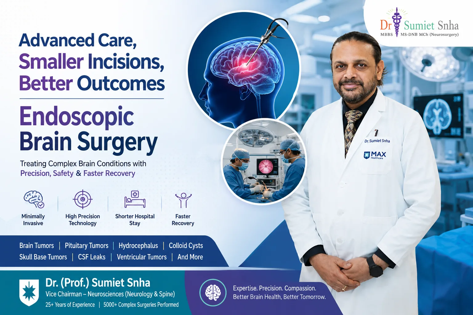
- One of the most experienced minimally invasive and endoscopic neurosurgeons in Delhi, with extensive experience in ETV and neuroendoscopic procedures.
- Former Professor of Neurosurgery at AIIMS, New Delhi, treating hydrocephalus in newborns, children, adults and the elderly.
- Expertise in the full range of CSF diversion - ETV, programmable shunts, shunt revisions and NPH evaluation.
- Paediatric and adult neuro-ICU, neonatal care and rehabilitation support at Max Super Speciality Hospital, Dwarka.
Cost Factors for Hydrocephalus Surgery
- Type of procedure - ETV, shunt or combined procedures.
- Type of shunt valve - fixed or programmable.
- Age of the patient and need for neonatal or paediatric ICU.
- Underlying cause, such as a tumor needing treatment.
- Pre-operative tests, such as lumbar drainage trials for NPH.
- Hospital stay and insurance coverage.
Living Well With Treated Hydrocephalus
Most children and adults with treated hydrocephalus lead active, fulfilling lives - attending school, working, playing sports within guidance, travelling and starting families. The keys are awareness and follow-up. Keep a record of the type of treatment (ETV or shunt), the valve model and settings, and share it with any doctor you see. Carry a medical information card. Teachers and caregivers should know the warning signs of shunt malfunction or ETV failure. Children with hydrocephalus sometimes have specific learning difficulties, such as problems with attention, memory or visual-spatial skills, even when their hydrocephalus is well controlled; early educational assessment and support can help them reach their full potential. With good care, the outlook for people with hydrocephalus today is far brighter than it was a generation ago.
Hydrocephalus Detected Before Birth
With routine prenatal ultrasound, enlarged ventricles (ventriculomegaly) are sometimes detected in an unborn baby. This naturally causes great anxiety for expectant parents. It is important to know that mild ventriculomegaly is relatively common and that many babies with mild enlargement are born healthy and develop normally. More significant enlargement may be associated with conditions such as aqueductal stenosis, spina bifida or other brain malformations. A detailed fetal scan, fetal MRI and, where appropriate, genetic testing help clarify the cause and outlook. Prenatal consultation with a neurosurgeon allows parents to understand what treatment might be needed after birth and to plan delivery at a centre with neonatal intensive care and neurosurgical facilities. After birth, the baby is examined and scanned, and treatment - if needed - is timed according to the baby's condition, weight and the cause of hydrocephalus.
Hydrocephalus in Premature Babies
Premature infants, particularly those born very early, are at risk of bleeding into the ventricles of the brain in the first days of life. In some babies, this blood blocks fluid pathways or impairs absorption, causing post-haemorrhagic hydrocephalus. Because these babies are small and fragile, a permanent shunt is often delayed until they are larger and the blood has cleared. In the meantime, temporary measures such as a ventricular access device (a small reservoir under the scalp from which fluid can be withdrawn) or a ventriculo-subgaleal shunt may be used to control pressure. Close cooperation between neonatologists and neurosurgeons is essential, and careful monitoring of head size and ultrasound findings guides decisions.
Understanding the Long-Term Outlook
The outlook for people with hydrocephalus depends largely on the underlying cause, how early treatment was provided, and whether there are associated brain conditions. Many children treated for aqueductal stenosis grow up with normal intelligence and lead entirely independent lives. Children whose hydrocephalus followed severe prematurity, infection or complex malformations may have additional challenges that require ongoing support. Adults treated for obstructive hydrocephalus due to tumors or cysts usually recover well once the cause is addressed, and older adults with NPH often regain walking ability and independence. Regardless of the cause, regular follow-up, prompt attention to warning signs and support for learning and development give each patient the best possible future.
Glossary
- Cerebrospinal fluid (CSF): the clear fluid that surrounds and cushions the brain and spinal cord.
- Ventricles: fluid-filled chambers within the brain.
- Aqueductal stenosis: narrowing of the channel between the third and fourth ventricles.
- ETV: endoscopic third ventriculostomy - creating an opening in the floor of the third ventricle.
- VP shunt: ventriculoperitoneal shunt - a tube draining CSF from the brain to the abdomen.
- Programmable valve: a shunt valve whose pressure setting can be adjusted externally.
- Papilloedema: swelling of the optic nerve head due to raised pressure in the head.
Beyond ETV - Other Neuroendoscopic Procedures for CSF Problems
The same slim endoscope used for third ventriculostomy can treat several other disorders of fluid circulation within the brain. These procedures share the benefits of a small burr hole, a short operation and, very often, avoiding a shunt or reducing the number of shunts a patient needs. Whether an endoscopic procedure is suitable depends on the detailed anatomy seen on MRI, including the size of the ventricles, the thickness of membranes and the position of blood vessels.
Arachnoid cyst fenestration
Arachnoid cysts are fluid-filled sacs lined by the brain's delicate arachnoid membrane. Most are found incidentally and need no treatment. When a cyst enlarges, compresses the brain or blocks fluid pathways - causing headaches, an enlarging head in infants, or hydrocephalus - the endoscope can be used to open its walls into the neighbouring ventricle or fluid spaces so that it drains naturally.
Septostomy and treatment of isolated ventricles
After infection or bleeding, membranes may divide the ventricles into separate compartments that each enlarge. Opening the septum pellucidum or these membranes endoscopically can connect the compartments so that a single drainage route, or a single shunt catheter, can treat them all.
Aqueductoplasty
In selected patients with a short narrowing of the aqueduct, particularly when the fourth ventricle is trapped, the narrowed channel itself may be carefully widened and sometimes stented. This is done only when the anatomy is favourable, as the area around the aqueduct is delicate.
Replacing a Failed Shunt With an ETV
Some people who have lived with a shunt for years face repeated blockages or infections. In a proportion of these patients - especially those whose hydrocephalus was originally due to an obstruction such as aqueductal stenosis or a tumour - the anatomy may now be suitable for an ETV. When a shunt malfunctions, an ETV may be performed at the time of revision, and in some cases the old shunt can be tied off or later removed, freeing the patient from dependence on the device.
This is not possible for everyone. The ventricles must be large enough to navigate safely, the floor of the third ventricle must be accessible, and the absorption pathways around the brain must be working. Careful MRI assessment helps predict suitability, and patients are monitored closely afterwards because the brain may take time to adjust to the new route of drainage. Where ETV is not appropriate, a well-planned shunt revision, sometimes with a programmable valve, remains a reliable solution.
Preparing for Hydrocephalus Surgery
Before surgery, the team reviews recent scans, checks blood counts and clotting, and assesses the patient's fitness for anaesthesia. Families should share details of any previous shunt, including the valve type and setting, operation notes and earlier scans, since comparing images over time is very helpful. Fasting instructions must be followed carefully. Patients from outside Delhi can send reports by WhatsApp on +91-8448877746 or email sumitneuro@gmail.com for a preliminary opinion, and video consultation is available. For an in-person visit at Max Super Speciality Hospital, Dwarka, from Monday to Saturday, please book an appointment. Children's care is described further on our paediatric neurosurgery page.
Frequently Asked Questions
Can hydrocephalus be cured?
Hydrocephalus can be effectively treated. ETV can provide a long-term solution without an implant in suitable patients, and shunts control hydrocephalus in most others, though they may need revision over time.
What is the success rate of ETV?
Success depends mainly on the cause and the patient's age. It is highest in older children and adults with obstructive hydrocephalus, such as aqueductal stenosis, and lower in young infants.
Will my child need a shunt for life?
Many people with shunts need them long term. In some cases, a shunt can later be replaced by an ETV if the anatomy is suitable.
How do I know if a shunt is blocked?
Symptoms include headache, vomiting, drowsiness, irritability and vision changes; in infants, a bulging fontanelle. Seek urgent medical care if these occur.
Can older adults benefit from hydrocephalus surgery?
Yes. Older adults with normal pressure hydrocephalus often show significant improvement in walking and bladder control after a shunt, especially when selected with a positive tap test.
Is ETV surgery painful?
It is performed under general anaesthesia through a small incision. Post-operative discomfort is usually mild and managed with simple pain relief.
Can I have an MRI with a programmable shunt?
Yes, but the valve setting may change with the magnetic field and should be checked and reset after the scan if needed. Some newer valves are resistant to such changes.
Expert Help for Hydrocephalus
Whether you are a parent worried about your baby's head growth, or an older adult noticing walking and memory changes, a specialist evaluation can provide answers and effective treatment. Book a consultation with Dr. (Prof.) Sumiet Snha at Max Hospital, Dwarka, New Delhi. Related pages: paediatric neurosurgery, brain tumor surgery and vascular neurosurgery.