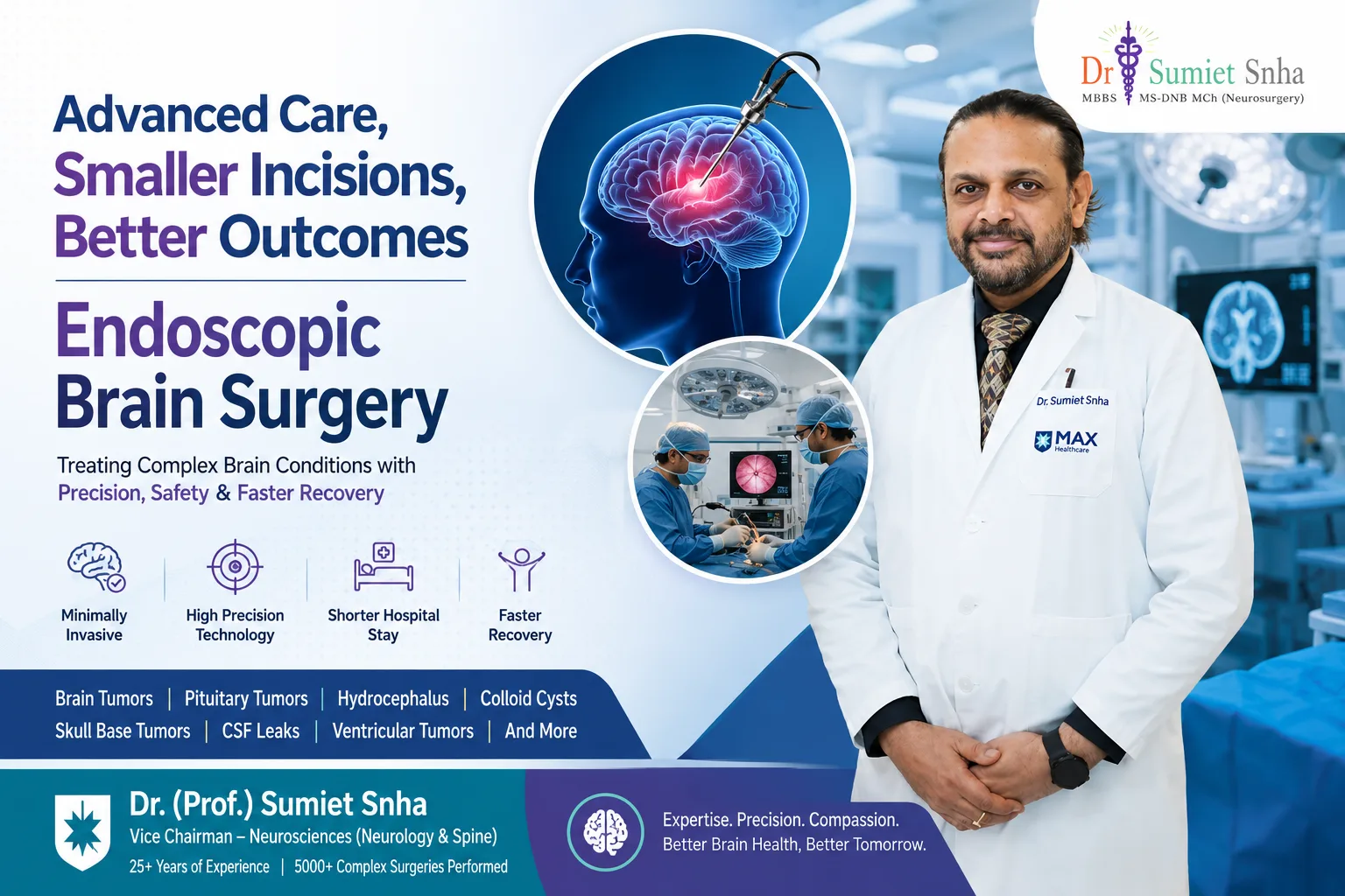
Brain surgery has advanced significantly over the past few decades, making many procedures safer, more precise, and less invasive than ever before. Endoscopic Brain Surgery is one of the most important innovations in modern neurosurgery, allowing surgeons to treat selected brain conditions through small openings using a high-definition camera (endoscope) and specially designed surgical instruments. This minimally invasive brain surgery technique helps access delicate areas of the brain while reducing disruption to surrounding healthy tissue.
Today, many patients and healthcare professionals prefer endoscopic neurosurgery because it often offers several advantages over conventional open brain surgery, including smaller incisions, reduced blood loss, less postoperative discomfort, shorter hospital stays, and faster recovery. Continuous advancements in surgical imaging, neuronavigation systems, and high-definition endoscopic technology have further improved the precision, safety, and overall success of brain endoscopy surgery for carefully selected patients.
Whether you have been diagnosed with a brain tumor, pituitary tumor, hydrocephalus, or another neurological condition, understanding your treatment options is essential for making informed decisions. In this complete patient guide, you'll learn how endoscopic brain surgery is performed, when it is recommended, its benefits and potential risks, what to expect during recovery, and who may be a suitable candidate for this advanced minimally invasive neurosurgery approach. The goal is to provide clear, evidence-based information that helps patients and their families better understand this modern surgical technique and discuss appropriate treatment options with their neurosurgical team.
What Is Endoscopic Brain Surgery?
Endoscopic Brain Surgery is a modern minimally invasive brain surgery technique that allows neurosurgeons to diagnose and treat certain brain disorders through very small openings or natural pathways instead of performing a large skull opening. The procedure uses a thin, flexible tube called an endoscope, which contains a high-definition camera and a powerful light source. This camera transmits magnified, real-time images of the surgical area to a monitor, allowing the surgeon to perform highly precise movements using specialized microsurgical instruments. Advances in endoscopic technology have expanded its role in modern endoscopic neurosurgery, making it an effective treatment option for carefully selected brain conditions.
How Does an Endoscope Work?
An endoscope is designed to provide excellent visualization of deep and delicate areas of the brain that may be difficult to access using conventional techniques. During brain endoscopy surgery, the neurosurgeon gently inserts the endoscope through a small incision in the skull or, in selected cases, through the nasal passages using an endoscopic endonasal approach. The high-definition camera delivers clear, magnified images that help the surgical team identify important nerves, blood vessels, and brain structures while minimizing injury to healthy tissue. Specialized instruments passed alongside the endoscope are then used to remove tumors, drain cysts, restore normal cerebrospinal fluid flow, or perform other necessary procedures with greater precision.
Endoscopic Brain Surgery vs. Traditional Open Brain Surgery
Traditional open brain surgery (craniotomy) usually requires a larger scalp incision and a wider opening in the skull to provide direct access to the brain. While it remains the preferred treatment for many complex neurological conditions, it may involve greater tissue exposure and a longer recovery period.
In contrast, Endoscopic Brain Surgery focuses on reaching the affected area through much smaller surgical openings or natural anatomical pathways whenever appropriate. Because the surgeon works with a magnified endoscopic view, there is often less disruption to surrounding healthy brain tissue. Many patients experience reduced postoperative pain, lower blood loss, shorter hospital stays, and an earlier return to daily activities. However, this technique is not suitable for every patient, and the choice between endoscopic and open surgery depends on factors such as the size, location, and complexity of the brain condition.
Advantages of Minimally Invasive Endoscopic Brain Surgery
The growing popularity of minimally invasive neurosurgery is largely due to its patient-centered benefits. Depending on the condition being treated, potential advantages may include:
- Smaller surgical incisions and minimal scarring
- Better visualization of deep brain structures through high-definition imaging
- Reduced trauma to healthy brain tissue
- Lower blood loss during surgery
- Reduced risk of infection in appropriately selected cases
- Less postoperative discomfort
- Shorter hospital stay
- Faster recovery and earlier return to normal activities
- Improved cosmetic outcomes
These benefits have made Endoscopic Brain Surgery an important advancement in modern neurosurgical care, although the most appropriate surgical approach should always be determined after a detailed neurological evaluation and imaging studies.
Common Conditions Treated with Endoscopic Brain Surgery
Today, Endoscopic Brain Surgery is used to manage a wide range of carefully selected neurological conditions. Common indications include:
- Brain tumors located in accessible areas
- Pituitary tumors
- Hydrocephalus, including endoscopic third ventriculostomy (ETV)
- Colloid cysts of the third ventricle
- Certain skull base tumors
- Intraventricular tumors
- Cerebrospinal fluid (CSF) leaks
- Selected brain cysts and other structural abnormalities
Ongoing improvements in surgical optics, neuronavigation, and specialized endoscopic instruments continue to expand the role of brain endoscopy surgery. Even so, not every brain disorder can be treated using an endoscopic approach. The decision depends on the patient's symptoms, imaging findings, overall health, and the expertise of the neurosurgical team. A personalized treatment plan remains the foundation of achieving the safest and most effective outcome.
When Is Endoscopic Brain Surgery Recommended?
Endoscopic Brain Surgery is recommended for carefully selected patients with brain conditions that can be safely treated using a minimally invasive brain surgery approach. Instead of making a large opening in the skull, this advanced technique allows neurosurgeons to access certain areas of the brain through small incisions or natural anatomical pathways with the help of a high-definition endoscope and specialized surgical instruments. However, not every neurological disorder is suitable for this procedure. The decision depends on the location, size, complexity of the condition, the patient's overall health, and detailed brain imaging such as MRI or CT scans.
Brain Tumors
Certain brain tumors can be treated using Endoscopic Brain Surgery, particularly when they are located within the brain's ventricular system or near the skull base. The endoscope provides a magnified view of the surgical field, allowing the neurosurgeon to remove or biopsy the tumor while minimizing disruption to surrounding healthy brain tissue. This brain tumor surgery approach may help reduce postoperative discomfort and support a faster recovery in appropriately selected patients.
Pituitary Tumors
One of the most common applications of endoscopic neurosurgery is the treatment of pituitary tumors. Many pituitary adenomas can be removed through an endoscopic endonasal approach, where the surgeon accesses the tumor through the nasal passages without making a visible external incision. This minimally invasive technique provides excellent visualization of the pituitary gland while reducing tissue trauma and shortening recovery time for many patients.
Hydrocephalus
Patients with hydrocephalus, a condition caused by excess cerebrospinal fluid (CSF) in the brain, may benefit from Endoscopic Brain Surgery. A procedure called Endoscopic Third Ventriculostomy (ETV) creates a small opening within the brain's ventricular system, allowing CSF to flow more naturally and reducing pressure inside the skull. In selected patients, ETV may serve as an effective alternative to long-term shunt placement.
Colloid Cysts
Colloid cysts are rare, fluid-filled cysts that commonly develop in the third ventricle of the brain. Although benign, they can block the normal circulation of cerebrospinal fluid, causing headaches, nausea, memory problems, or hydrocephalus. Brain endoscopy surgery allows many of these cysts to be removed through a minimally invasive approach, helping restore normal CSF flow while reducing surgical trauma.
Skull Base Tumors
Several carefully selected skull base tumors can be treated using Endoscopic Brain Surgery, particularly those located around the pituitary gland, sphenoid sinus, or anterior skull base. The endoscopic approach provides enhanced visualization of deep anatomical structures while avoiding large external incisions. This technique has become an important option in modern minimally invasive neurosurgery for selected skull base conditions.
Ventricular Tumors
Tumors located inside the brain's ventricular system can often be challenging to access through conventional surgery. In selected cases, endoscopic brain tumor surgery allows neurosurgeons to obtain tissue samples (biopsy), remove smaller tumors, or relieve obstruction of cerebrospinal fluid pathways using specialized endoscopic instruments. The suitability of this technique depends on the tumor's size, location, and characteristics.
Cerebrospinal Fluid (CSF) Leaks
A cerebrospinal fluid (CSF) leak occurs when the protective fluid surrounding the brain escapes through a defect in the skull base. Persistent CSF leaks increase the risk of serious infections such as meningitis. Many skull base CSF leaks can now be repaired using endoscopic skull base surgery, which enables surgeons to seal the defect through the nasal passages without the need for traditional open surgery.
Selected Vascular and Neurological Disorders
Although conventional microsurgery remains the standard treatment for many vascular brain conditions, Endoscopic Brain Surgery may assist in selected cases involving certain vascular abnormalities, intracranial cysts, ventricular disorders, and other neurological conditions. In some procedures, the endoscope is used alongside microscopic techniques to improve visualization and surgical precision.
Symptoms That May Require Evaluation by a Neurosurgeon
Persistent neurological symptoms should never be ignored, as they may indicate an underlying brain condition requiring expert assessment. Patients should consult a neurosurgeon if they experience:
- Persistent or severe headaches
- Recurrent seizures
- Vision changes or double vision
- Progressive weakness or numbness in the face, arms, or legs
- Difficulty with balance or walking
- Persistent nausea or vomiting without a clear cause
- Memory problems or personality changes
- Difficulty speaking or understanding speech
- Symptoms of increased pressure inside the skull
- Fluid leaking from the nose after head injury or surgery
These symptoms do not always mean surgery is required, but they should be evaluated promptly through a neurological examination and appropriate imaging studies. Early diagnosis allows specialists to determine whether Endoscopic Brain Surgery, another form of brain tumor treatment, or a non-surgical approach is the safest and most effective option for each individual patient. A personalized treatment plan developed by an experienced neurosurgical team remains essential for achieving the best possible outcomes
How Endoscopic Brain Surgery Is Performed: Step-by-Step Procedure
Understanding how Endoscopic Brain Surgery is performed can help patients feel more informed and confident before treatment. This advanced minimally invasive brain surgery technique follows a carefully planned process that combines detailed preoperative evaluation, modern imaging technology, high-definition endoscopic visualization, and specialized microsurgical instruments. Every procedure is personalized according to the patient's diagnosis, the location of the brain lesion, and overall health. Although the exact surgical approach may vary depending on the condition being treated, the following steps explain how brain endoscopy surgery is typically performed.
Preoperative Evaluation and Diagnostic Imaging
Before recommending Endoscopic Brain Surgery, the neurosurgical team performs a comprehensive medical evaluation to confirm the diagnosis and determine whether the patient is an appropriate candidate for a minimally invasive approach.
The evaluation usually includes:
- Detailed medical history and physical examination
- Comprehensive neurological examination
- Magnetic Resonance Imaging (MRI)
- Computed Tomography (CT) scan
- Blood investigations and routine pre-anesthetic tests
- Additional imaging such as MR angiography or CT angiography when required
MRI provides highly detailed images of the brain, helping surgeons identify the exact size, location, and relationship of the abnormality to nearby nerves and blood vessels. CT scans help evaluate the skull and detect bleeding or calcification. These imaging studies allow the neurosurgical team to create a precise surgical plan while minimizing risks during endoscopic neurosurgery.
Administration of General Anesthesia
On the day of surgery, patients are taken to the operating room where an experienced anesthesiology team administers general anesthesia. This ensures the patient remains asleep, pain-free, and completely still throughout the procedure.
During surgery, vital signs including heart rate, blood pressure, oxygen levels, body temperature, and brain function are continuously monitored. Careful anesthetic management contributes significantly to patient safety during brain surgery procedures.
Making a Small Surgical Incision
Once the patient is safely under anesthesia, the neurosurgeon makes a carefully planned small incision. The location of the incision depends on the specific brain condition being treated.
For certain skull base or pituitary tumors, the surgeon may reach the surgical site through the natural nasal passages using an endoscopic endonasal approach, eliminating the need for an external incision. For other conditions, a small incision is made on the scalp to provide access to the affected area while preserving as much healthy tissue as possible.
Creating a Tiny Opening in the Skull (When Required)
If the condition cannot be accessed through natural pathways, the surgeon creates a very small opening in the skull, commonly called a burr hole or mini-craniotomy.
Unlike traditional open brain surgery, this opening is significantly smaller and is designed to provide sufficient access for the endoscope and surgical instruments. The minimally invasive approach helps reduce tissue disruption while maintaining excellent surgical visibility.
Inserting the Endoscope
The next step involves carefully inserting the endoscope, a thin tube equipped with a high-definition camera and a powerful light source.
As the endoscope advances toward the treatment area, it transmits magnified real-time images to large surgical monitors. This enhanced visualization allows the neurosurgeon to navigate delicate brain structures with exceptional accuracy while avoiding unnecessary injury to surrounding healthy tissue.
This advanced imaging capability is one of the major reasons why Endoscopic Brain Surgery has become an important development in minimally invasive neurosurgery.
Real-Time Visualization Using High-Definition Imaging
One of the greatest advantages of brain endoscopy surgery is the ability to obtain highly magnified, illuminated views of structures that are difficult to visualize during conventional surgery.
Modern endoscopic systems often include:
- High-definition (HD) cameras
- 4K visualization technology
- Angled endoscopes for improved viewing around corners
- Image-guided neuronavigation systems
- Advanced surgical microscopes when additional visualization is required
These technologies enable surgeons to identify tumors, cysts, blood vessels, nerves, and surrounding brain tissue with remarkable precision, supporting safer surgical decision-making throughout the procedure.
Removal or Treatment of the Affected Tissue
After reaching the target area, the neurosurgeon introduces specialized microsurgical instruments alongside the endoscope.
Depending on the patient's condition, the surgeon may:
- Remove a brain tumor
- Excise a colloid cyst
- Remove or biopsy a ventricular lesion
- Treat a pituitary tumor
- Perform an Endoscopic Third Ventriculostomy (ETV) for hydrocephalus
- Repair a cerebrospinal fluid (CSF) leak
- Restore normal cerebrospinal fluid circulation
Throughout the operation, every movement is performed under continuous visual guidance to maximize surgical precision while protecting important neurological structures. The primary goal is to treat the disease effectively while preserving normal brain function whenever possible.
Bleeding Control and Surgical Closure
Once the surgical objective has been achieved, the neurosurgeon carefully inspects the operative field to ensure complete bleeding control.
Specialized techniques are used to seal small blood vessels, reduce the risk of postoperative bleeding, and verify that surrounding structures remain intact. After removing the endoscope and surgical instruments, the small skull opening is secured when necessary, and the incision is closed using fine sutures or surgical staples.
Because Endoscopic Brain Surgery requires much smaller incisions than conventional procedures, many patients experience less postoperative discomfort and improved cosmetic healing.
Postoperative Monitoring and Recovery Unit Care
Following surgery, patients are transferred to the recovery room or neuro-intensive care unit for close observation.
During this period, healthcare professionals monitor:
- Neurological function
- Blood pressure and heart rate
- Oxygen levels
- Level of consciousness
- Vision and speech
- Limb strength and movement
- Pain control
- Signs of bleeding or infection
Depending on the type of surgery and the patient's recovery, additional MRI or CT scans may be performed to evaluate the surgical outcome. Many patients undergoing minimally invasive brain surgery are able to begin gentle movement earlier and may have shorter hospital stays compared with traditional open procedures, although recovery varies based on the underlying condition and the complexity of the surgery.
Personalized Surgical Planning Is Essential
Although Endoscopic Brain Surgery offers many advantages, every patient's condition is unique. Factors such as the size and location of the lesion, overall health, previous surgeries, and neurological status determine whether this approach is appropriate. An experienced neurosurgeon will carefully evaluate these factors and recommend the safest, most effective treatment plan to achieve the best possible outcome while minimizing surgical risks.
Benefits of Endoscopic Brain Surgery
Endoscopic Brain Surgery has transformed modern neurosurgery by offering a minimally invasive brain surgery approach for selected brain disorders. Unlike traditional open brain surgery, this advanced technique uses a high-definition endoscope and specialized microsurgical instruments to reach the affected area through much smaller openings or natural anatomical pathways. As a result, many patients experience a smoother recovery while maintaining excellent surgical outcomes. However, the benefits depend on the patient's diagnosis, the location of the brain lesion, and careful patient selection by an experienced neurosurgeon.
Smaller Incision
One of the most noticeable advantages of Endoscopic Brain Surgery is the use of a much smaller surgical incision compared with conventional craniotomy. In some procedures, such as endoscopic skull base surgery for pituitary tumors, surgeons can even access the brain through the nasal passages without making an external incision. Smaller incisions generally lead to less tissue disruption, reduced scarring, and improved patient comfort during recovery.
Less Damage to Healthy Brain Tissue
Modern endoscopic neurosurgery provides surgeons with a magnified, high-definition view of delicate brain structures. This enhanced visualization allows the surgical team to work with greater precision while minimizing unnecessary manipulation of healthy brain tissue. Preserving normal brain structures is particularly important because it may help maintain neurological function and reduce postoperative complications.
Reduced Blood Loss
Because brain endoscopy surgery requires smaller surgical openings and precise instrument handling, many procedures are associated with reduced blood loss compared with traditional open brain surgery. Less bleeding may decrease the need for blood transfusions and contribute to a safer surgical experience for appropriately selected patients.
Lower Risk of Infection
Every surgical procedure carries some risk of infection, but minimally invasive techniques may help reduce that risk by limiting the size of the incision and minimizing exposure of brain tissue. Smaller wounds are generally easier to heal and require less extensive postoperative wound care. Strict sterile surgical protocols and experienced neurosurgical teams remain essential for preventing infection.
Less Postoperative Pain
Patients undergoing Endoscopic Brain Surgery often experience less postoperative discomfort because the procedure causes less disruption to the scalp, skull, muscles, and surrounding tissues. Although pain levels vary from person to person, many patients require less pain medication and become comfortable more quickly than after traditional open brain surgery.
Shorter Hospital Stay
A major benefit of minimally invasive brain surgery is the potential for a shorter hospital stay. Since surgical trauma is reduced and recovery often begins earlier, many patients can return home sooner once their neurological condition is stable. The length of hospitalization depends on the underlying brain condition, the complexity of the surgery, and the patient's overall recovery.
Faster Recovery
Recovery following Endoscopic Brain Surgery is often quicker than recovery after conventional open procedures. Smaller incisions, reduced tissue trauma, and lower postoperative discomfort may allow patients to regain strength, resume light activities, and begin rehabilitation earlier. Nevertheless, recovery time varies depending on the type of surgery performed and each patient's overall health.
Improved Cosmetic Results
Cosmetic outcomes are another important advantage of brain endoscopy surgery. Because the surgical incisions are smaller—and in some skull base procedures completely hidden inside the nose—patients usually have minimal visible scarring. This can improve confidence and overall satisfaction after treatment while supporting excellent functional outcomes.
Earlier Return to Normal Activities
Many patients who undergo Endoscopic Brain Surgery are able to return to their normal daily activities, work, and social life sooner than those recovering from larger open brain procedures. The exact timeline depends on the medical condition being treated, neurological recovery, and the surgeon's recommendations. Following postoperative instructions, attending follow-up appointments, and completing rehabilitation when required all contribute to a safe and successful recovery.
Why Many Patients Choose Endoscopic Brain Surgery
The growing popularity of Endoscopic Brain Surgery reflects significant advances in minimally invasive neurosurgery, surgical imaging, neuronavigation systems, and high-definition endoscopic technology. For carefully selected patients, this approach offers precise treatment with smaller incisions, less damage to healthy brain tissue, reduced blood loss, less postoperative pain, shorter hospital stays, and faster recovery. However, it is important to remember that not every brain condition is suitable for an endoscopic approach. A thorough neurological evaluation, detailed MRI or CT imaging, and consultation with an experienced neurosurgeon are essential to determine the safest and most effective treatment plan for each individual patient.
Risks and Possible Complications of Endoscopic Brain Surgery
Like all neurosurgical procedures, Endoscopic Brain Surgery carries certain risks and potential complications. Although this minimally invasive brain surgery technique is designed to reduce surgical trauma and improve recovery, no operation is completely risk-free. The likelihood of complications depends on several factors, including the patient's overall health, the type and location of the brain condition, the complexity of the procedure, and the experience of the neurosurgical team. Before surgery, your neurosurgeon will carefully explain the potential benefits and risks to help you make an informed treatment decision.
Bleeding
Bleeding is one of the possible complications of any brain surgery procedure. Although Endoscopic Brain Surgery generally involves smaller incisions and less tissue disruption than traditional open surgery, bleeding can still occur during or after the operation. Neurosurgeons use advanced microsurgical techniques, specialized instruments, and continuous monitoring to control bleeding and minimize this risk. In rare situations, additional treatment may be required if significant bleeding develops.
Infection
Despite strict sterile protocols in the operating room, infection remains a potential complication of brain endoscopy surgery. Infections may occur at the surgical incision, within the skull, or, rarely, around the brain's protective tissues. To reduce this risk, patients receive appropriate preventive measures, including careful skin preparation, sterile surgical techniques, and antibiotics when indicated. Prompt medical attention is important if fever, increasing wound redness, swelling, or drainage develops after surgery.
Cerebrospinal Fluid (CSF) Leakage
A cerebrospinal fluid (CSF) leak occurs when the protective fluid surrounding the brain escapes through a small opening in the membranes covering the brain. This complication is more commonly associated with endoscopic skull base surgery, particularly when treating pituitary tumors or repairing skull base defects. Symptoms may include clear fluid leaking from the nose, persistent headaches, or signs of infection. Modern surgical closure techniques have significantly reduced the incidence of CSF leaks, and most cases can be successfully managed when detected early.
Injury to Nearby Nerves or Brain Tissue
The brain contains delicate nerves and vital structures responsible for movement, speech, vision, memory, balance, and other essential functions. Although Endoscopic Brain Surgery offers excellent magnified visualization, there remains a small possibility of injury to nearby nerves or healthy brain tissue during complex procedures. The exact risk depends on the size and location of the lesion being treated. Advanced neuronavigation systems, high-definition endoscopes, and experienced surgical techniques help reduce this possibility.
Temporary Neurological Symptoms
Some patients may experience temporary neurological symptoms following minimally invasive brain surgery. These symptoms vary depending on the surgical site and may include:
- Mild weakness
- Numbness or tingling
- Temporary vision changes
- Balance difficulties
- Headache
- Fatigue
- Short-term memory or concentration problems
In many cases, these symptoms gradually improve as the brain heals. Rehabilitation therapies such as physiotherapy, occupational therapy, or speech therapy may be recommended to support recovery when needed.
Anesthesia-Related Risks
Since Endoscopic Brain Surgery is usually performed under general anesthesia, there are potential risks related to anesthesia itself. Although serious complications are uncommon, possible issues may include allergic reactions, breathing difficulties, blood pressure changes, heart rhythm disturbances, nausea, vomiting, or temporary confusion after surgery. An experienced anesthesiology team carefully evaluates each patient's medical history and continuously monitors vital signs throughout the procedure to maximize safety.
Need for Additional Surgery in Some Cases
While Endoscopic Brain Surgery is highly effective for many carefully selected conditions, additional treatment or surgery may occasionally be necessary. This may happen if:
- Complete tumor removal is not safely possible.
- A tumor recurs over time.
- Persistent cerebrospinal fluid leakage develops.
- Bleeding or infection requires further management.
- The patient's condition changes during follow-up.
Your neurosurgeon will discuss these possibilities before surgery and explain whether additional treatment may be required as part of long-term care.
The Importance of Choosing an Experienced Neurosurgical Team
One of the most important factors influencing the success of Endoscopic Brain Surgery is the experience and expertise of the neurosurgical team. These procedures require specialized training, advanced surgical skills, and access to modern technology such as high-definition endoscopes, neuronavigation systems, intraoperative imaging, and specialized microsurgical instruments.
When selecting a neurosurgeon and hospital, patients should consider:
- Experience in endoscopic neurosurgery
- Expertise in treating complex brain disorders
- Availability of advanced surgical technology
- Comprehensive neurocritical care facilities
- Multidisciplinary team support
- Proven commitment to patient safety and evidence-based care
Balancing Benefits and Risks
Although every surgical procedure carries potential risks, Endoscopic Brain Surgery has become an important advancement in modern minimally invasive neurosurgery because it often reduces tissue damage, blood loss, postoperative pain, and recovery time compared with conventional open brain surgery. Careful patient selection, detailed preoperative planning, advanced imaging, and experienced surgical care help maximize the benefits while minimizing complications. Patients should discuss all available treatment options, expected outcomes, and potential risks with their neurosurgeon to make an informed decision based on their individual condition.
Recovery After Endoscopic Brain Surgery
Recovery after Endoscopic Brain Surgery is generally faster than recovery from traditional open brain surgery because this minimally invasive brain surgery technique uses smaller incisions and causes less disruption to healthy brain tissue. However, every patient's recovery is unique and depends on the condition being treated, the complexity of the procedure, age, overall health, and adherence to postoperative instructions. Understanding the recovery process helps patients and their families prepare for a safe and successful return to daily life.
Recovery Timeline After Endoscopic Brain Surgery
|
Recovery Stage |
What to Expect |
|
First 24 Hours |
Close monitoring in the recovery room or Neuro ICU, neurological assessments, pain control, and vital sign monitoring. |
|
Hospital Stay |
Usually 2–5 days for uncomplicated cases, although some patients may require a longer stay depending on their condition. |
|
First 2 Weeks |
Rest, wound care, gradual walking, light daily activities, and follow-up consultation with the neurosurgeon. |
|
2–6 Weeks |
Progressive improvement in energy levels, reduced discomfort, and gradual return to routine activities as advised by the medical team. |
|
6–12 Weeks |
Many patients can return to work and resume most normal activities, depending on the type of surgery and their recovery progress. |
Immediate Postoperative Care
After Endoscopic Brain Surgery, patients are transferred to the recovery room or Neuro Intensive Care Unit (Neuro ICU), where healthcare professionals closely monitor neurological function, blood pressure, heart rate, oxygen levels, vision, speech, movement, and level of consciousness. Regular neurological examinations help detect any early complications and ensure the brain is recovering as expected.
Hospital Stay
One of the advantages of brain endoscopy surgery is a shorter hospital stay compared with many traditional brain surgeries. Most patients remain hospitalized for observation, pain management, and monitoring until they are medically stable. Before discharge, the neurosurgical team provides detailed instructions regarding medications, wound care, diet, and follow-up appointments.
Pain Management
Although Endoscopic Brain Surgery generally causes less postoperative pain, mild headaches, discomfort around the incision, fatigue, or nasal congestion (after endoscopic endonasal surgery) are common during the early recovery period. Pain is usually managed with prescribed medications, adequate hydration, and sufficient rest. Patients should take medicines only as directed by their treating physician.
Activity Restrictions
During the first few weeks after minimally invasive brain surgery, patients should avoid:
- Heavy lifting
- Strenuous exercise
- Driving until medically cleared
- Smoking and alcohol consumption
- Activities that increase pressure inside the head
Light walking is usually encouraged because it improves circulation, reduces the risk of blood clots, and supports overall recovery.
Follow-Up Imaging
Follow-up care is an important part of recovery after Endoscopic Brain Surgery. Depending on the underlying condition, the neurosurgeon may recommend MRI or CT scans to evaluate healing, confirm successful treatment, and monitor for recurrence or complications. Regular follow-up visits also allow the healthcare team to assess neurological recovery and answer any patient concerns.
Rehabilitation When Needed
Some patients recover without additional therapy, while others may benefit from rehabilitation depending on the brain condition treated. Rehabilitation programs may include:
- Physiotherapy to improve strength, balance, and mobility
- Occupational therapy to regain independence in daily activities
- Speech and language therapy if speech or swallowing has been affected
- Neuropsychological support for memory, concentration, or cognitive rehabilitation
A personalized rehabilitation plan helps maximize recovery and improve long-term quality of life.
Warning Signs That Require Immediate Medical Attention
Patients should contact their neurosurgeon or seek emergency medical care immediately if they experience:
- Severe or worsening headache
- Persistent vomiting
- High fever or chills
- Clear fluid leaking from the nose or surgical wound
- Increasing redness, swelling, or discharge from the incision
- New weakness or numbness
- Difficulty speaking or understanding speech
- Vision changes
- Seizures
- Confusion, excessive drowsiness, or loss of consciousness
Early recognition and treatment of these symptoms can prevent serious complications and improve outcomes.
Returning to Work and Normal Activities
Most patients gradually return to work and regular daily activities within 4 to 12 weeks, depending on the type of Endoscopic Brain Surgery, the condition treated, and the physical demands of their occupation. Individuals with physically demanding jobs may require a longer recovery period than those performing desk-based work. Recovery should always progress according to the neurosurgeon's recommendations rather than a fixed timeline.
A Successful Recovery Requires Teamwork
Successful recovery after Endoscopic Brain Surgery depends not only on the procedure itself but also on careful postoperative care, regular follow-up visits, adherence to medications, healthy nutrition, adequate sleep, and gradual physical activity. By closely following medical advice and reporting any unusual symptoms promptly, patients can support optimal healing and safely return to their normal lifestyle after minimally invasive brain surgery.
Who Is a Good Candidate for Endoscopic Brain Surgery?
Endoscopic Brain Surgery is an advanced minimally invasive brain surgery technique that offers significant benefits for many patients. However, not everyone with a neurological condition is a suitable candidate for this approach. The decision to perform brain endoscopy surgery is based on a detailed medical evaluation, advanced imaging studies, and careful assessment by an experienced neurosurgical team. The primary goal is to choose the safest and most effective treatment that provides the best possible outcome while preserving normal brain function.
Factors Considered Before Surgery
Before recommending Endoscopic Brain Surgery, the neurosurgeon carefully reviews several medical and surgical factors. This evaluation helps determine whether the condition can be treated safely using a minimally invasive approach.
The assessment generally includes:
- Detailed medical history
- Comprehensive neurological examination
- MRI and CT scan findings
- Type of brain condition
- Severity of symptoms
- Patient's overall medical condition
- Previous treatments or surgeries
- Potential benefits and risks of the procedure
A personalized evaluation ensures that every treatment plan is tailored to the patient's specific needs rather than using a one-size-fits-all approach.
Tumor Size and Location
One of the most important factors influencing candidacy is the size and location of the brain lesion. Endoscopic Brain Surgery is most effective for carefully selected tumors and conditions located in areas that can be safely reached through a minimally invasive pathway.
Common conditions that may be suitable include:
- Pituitary tumors
- Colloid cysts
- Certain ventricular tumors
- Selected skull base tumors
- Hydrocephalus requiring Endoscopic Third Ventriculostomy (ETV)
- Some cerebrospinal fluid (CSF) leaks
Large, deeply invasive, highly vascular, or complex tumors involving multiple critical brain structures may require conventional microsurgery or a combination of surgical techniques to achieve the safest result.
Overall Health
A patient's general health plays a major role in determining whether minimally invasive neurosurgery is appropriate. Before surgery, doctors evaluate:
- Heart and lung function
- Blood pressure
- Diabetes control
- Kidney and liver function
- Blood clotting ability
- Current medications
- Presence of other medical conditions
Patients who are medically stable are generally better candidates because they are more likely to tolerate anesthesia well and recover smoothly after surgery.
Age Considerations
Age alone does not determine eligibility for Endoscopic Brain Surgery. Both children and older adults may benefit from brain endoscopy surgery when clinically appropriate. Instead of focusing only on age, neurosurgeons consider:
- Functional status
- Neurological condition
- Medical fitness
- Expected surgical benefits
- Recovery potential
Many elderly patients successfully undergo minimally invasive procedures when they are carefully selected and medically optimized before surgery.
Previous Brain Surgeries
Patients who have undergone previous brain surgery can still be considered for Endoscopic Brain Surgery, although additional planning may be required. Scar tissue, altered anatomy, implanted devices, or previous radiation treatment can influence the choice of surgical approach.
Advanced MRI, CT imaging, and neuronavigation systems help neurosurgeons determine whether an endoscopic technique remains the safest option or whether another surgical method would provide better access and lower risk.
When Traditional Open Brain Surgery May Be Recommended
Although Endoscopic Brain Surgery offers numerous advantages, it is not the best treatment for every patient. In some situations, traditional open brain surgery (craniotomy) provides better visualization, wider access, or greater surgical control.
Traditional surgery may be recommended when:
- The tumor is very large.
- The lesion involves multiple brain regions.
- There is significant bleeding requiring extensive control.
- Complete tumor removal cannot be safely achieved through an endoscope.
- Complex vascular abnormalities require open microsurgical treatment.
- Emergency surgery is needed after severe brain trauma.
The choice between endoscopic neurosurgery and conventional surgery is always based on patient safety, long-term neurological outcomes, and the nature of the underlying disease.
The Importance of Individualized Treatment Planning
Every neurological condition is unique, which is why individualized treatment planning is one of the most important aspects of modern neurosurgery. An experienced neurosurgeon carefully reviews imaging findings, symptoms, medical history, laboratory results, and patient expectations before recommending surgery.
A multidisciplinary team—including neurosurgeons, neuroradiologists, neuroanesthesiologists, neurologists, and rehabilitation specialists—often works together to develop a personalized treatment strategy. This collaborative approach helps determine whether Endoscopic Brain Surgery, another form of brain tumor surgery, or non-surgical management will provide the safest and most effective outcome.
Making the Right Treatment Decision
Choosing the appropriate surgical approach is a highly individualized decision that should never be based on a single factor. While Endoscopic Brain Surgery has revolutionized the treatment of many brain disorders through minimally invasive brain surgery, careful patient selection remains essential for achieving successful results. A thorough neurological evaluation, high-quality MRI and CT imaging, and consultation with an experienced endoscopic neurosurgery specialist help ensure that every patient receives the treatment best suited to their condition, maximizing safety, improving recovery, and supporting the best possible long-term neurological outcome.
Choosing the Right Neurosurgeon and Hospital
Selecting the right neurosurgeon and hospital is one of the most important decisions for anyone considering Endoscopic Brain Surgery. While this minimally invasive brain surgery technique offers many advantages, successful outcomes depend not only on advanced technology but also on the expertise of the neurosurgical team. Patients should look for a specialist with extensive experience in endoscopic neurosurgery, access to high-definition endoscopic systems, neuronavigation technology, modern operating theatres, and comprehensive neurocritical care facilities. A multidisciplinary approach involving neurologists, neuroradiologists, neuroanesthesiologists, rehabilitation specialists, and critical care experts ensures that every stage of treatment—from diagnosis to recovery—is carefully coordinated.
When evaluating a hospital, it is important to consider its infrastructure, infection control standards, availability of advanced imaging such as MRI and CT scans, intensive care support, and commitment to evidence-based treatment protocols. Hospitals that regularly perform complex brain endoscopy surgery procedures often have established systems for managing challenging neurological conditions and postoperative care.
An experienced neurosurgeon also understands that every patient requires an individualized treatment plan. Rather than recommending surgery for everyone, the best specialists carefully assess the patient's condition, explain all available treatment options, discuss expected outcomes and potential risks, and provide long-term follow-up care.
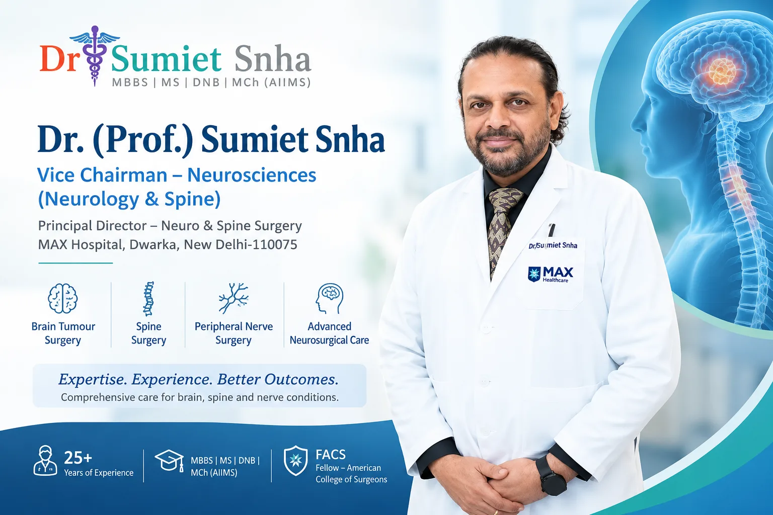
Dr. (Prof.) Sumiet Snha, Vice Chairman – Neurosciences (Neurology & Spine), has over 25+years of experience in neurosurgery and is dedicated to delivering patient-centered, evidence-based neurological care. His approach emphasizes accurate diagnosis, advanced surgical techniques, personalized treatment planning, and comprehensive follow-up to help patients achieve the best possible outcomes while prioritizing safety and quality of lif
Frequently Asked Questions
1. What is Endoscopic Brain Surgery?
Endoscopic Brain Surgery is a minimally invasive neurosurgical procedure that uses a thin, high-definition camera called an endoscope along with specialized surgical instruments to diagnose and treat selected brain conditions through small incisions or natural anatomical pathways. It helps improve surgical precision while minimizing damage to surrounding healthy brain tissue.
2. How is Endoscopic Brain Surgery different from open brain surgery?
Unlike traditional open brain surgery, which usually requires a larger incision and a wider opening in the skull, Endoscopic Brain Surgery uses much smaller openings. This often results in less tissue disruption, reduced blood loss, less postoperative pain, shorter hospital stays, and faster recovery for suitable patients.
3. Is Endoscopic Brain Surgery safe?
Yes. Endoscopic Brain Surgery is considered a safe and effective treatment for carefully selected patients when performed by an experienced neurosurgical team using modern imaging and surgical technology. As with any surgical procedure, there are potential risks, but careful planning and advanced techniques help minimize complications.
4. Which brain conditions can be treated with endoscopic surgery?
This technique is commonly used for selected brain tumors, pituitary tumors, hydrocephalus, colloid cysts, ventricular tumors, skull base tumors, cerebrospinal fluid (CSF) leaks, and certain other neurological conditions. Suitability depends on the size, location, and complexity of the condition.
5. How long does Endoscopic Brain Surgery usually take?
The duration of surgery varies depending on the condition being treated and its complexity. Most procedures take between 2 and 6 hours, although more complex cases may require additional operating time.
6. How many days do patients stay in the hospital after surgery?
Most patients remain in the hospital for approximately 2 to 5 days after uncomplicated Endoscopic Brain Surgery. However, the exact length of stay depends on the patient's neurological condition, recovery progress, and the type of surgery performed.
7. What is the recovery time after Endoscopic Brain Surgery?
Recovery varies for every patient. Many people return to light daily activities within 2 to 4 weeks, while returning to work or more strenuous activities may take 4 to 12 weeks, depending on the underlying condition and the neurosurgeon's advice.
8. Will there be a visible scar after surgery?
In most cases, scarring is minimal because Endoscopic Brain Surgery uses small incisions. For procedures performed through the nasal passages, such as endoscopic surgery for pituitary tumors, there is usually no visible external scar.
9. What are the possible risks and complications?
Although generally safe, potential risks include bleeding, infection, cerebrospinal fluid (CSF) leakage, temporary neurological symptoms, injury to nearby nerves or brain tissue, anesthesia-related complications, and, in some cases, the need for additional surgery. Your neurosurgeon will discuss these risks before treatment.
10. Can brain tumors be completely removed using Endoscopic Brain Surgery?
Some brain tumors can be completely removed using Endoscopic Brain Surgery, while others may require partial removal, biopsy, or a different surgical approach. The outcome depends on the tumor's size, location, type, and relationship to surrounding brain structures.
11. Who is eligible for Endoscopic Brain Surgery?
Patients with selected brain conditions that can be safely accessed using a minimally invasive approach may be suitable candidates. Eligibility is determined after a detailed neurological examination, MRI and CT imaging, review of overall health, and consultation with an experienced neurosurgeon.
12. How do I choose the right neurosurgeon for Endoscopic Brain Surgery?
Choose a neurosurgeon with extensive experience in endoscopic neurosurgery, access to advanced surgical technology, a multidisciplinary medical team, and a hospital equipped with modern neurocritical care facilities. It is also important to select a specialist who provides personalized treatment planning, explains all available treatment options, and offers comprehensive postoperative follow-up care.
Conclusion
Endoscopic Brain Surgery has significantly advanced the field of modern neurosurgery by making it possible to treat many brain disorders through minimally invasive brain surgery techniques. Using high-definition endoscopic technology and specialized surgical instruments, neurosurgeons can access certain areas of the brain with greater precision while minimizing disruption to healthy tissue. For many carefully selected patients, this approach offers important benefits, including smaller incisions, reduced blood loss, less postoperative pain, shorter hospital stays, improved cosmetic outcomes, and faster recovery compared with traditional open brain surgery.
However, Endoscopic Brain Surgery is not the right option for every patient or every neurological condition. The choice of treatment depends on factors such as the size and location of the brain lesion, the patient's overall health, detailed MRI and CT imaging findings, and the expertise of the treating neurosurgical team. A comprehensive neurological evaluation is essential to determine the safest and most effective treatment plan.
Persistent headaches, seizures, vision changes, weakness, numbness, balance problems, memory difficulties, or other unexplained neurological symptoms should never be ignored. Early diagnosis often leads to more treatment options and better outcomes. Consulting an experienced neurosurgeon such as Dr. (Prof.) Sumiet Snha, Vice Chairman – Neurosciences (Neurology & Spine), ensures that patients receive an accurate diagnosis, individualized treatment planning, and comprehensive follow-up care. A patient-centered approach combined with advanced neurosurgical techniques can help achieve the best possible clinical outcomes while supporting long-term brain health and quality of life.

0 Comments