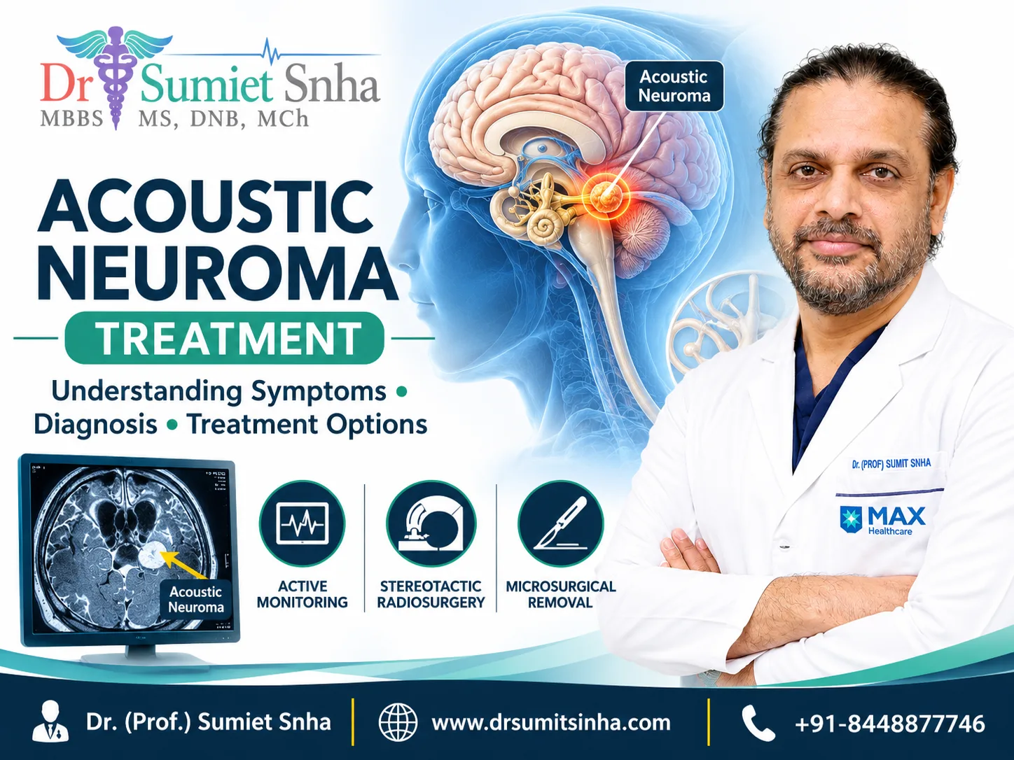
Acoustic Neuroma Treatment in Delhi involves evaluating a non-cancerous tumour that develops on the nerve responsible for transmitting hearing and balance signals from the inner ear to the brain. Although an acoustic neuroma usually grows slowly, its location near important nerves and the brainstem makes accurate diagnosis and appropriate treatment planning essential.
An acoustic neuroma is also known as a vestibular schwannoma. Some patients experience gradual hearing loss in one ear, ringing in the ear, dizziness or balance difficulties, while others may have few noticeable symptoms during the early stages.
Not every patient needs immediate surgery. Depending on the tumour's size, growth pattern, symptoms, hearing status and overall health, treatment may involve regular monitoring, stereotactic radiosurgery or microsurgical removal.
Understanding these options can help patients make informed decisions with their medical team. This guide explains the factors that influence treatment selection, the role of hearing preservation, possible complications and the importance of long-term follow-up.
What Is an Acoustic Neuroma (Vestibular Schwannoma)?
An acoustic neuroma is generally a benign, non-cancerous tumour that develops from Schwann cells surrounding the vestibular portion of the vestibulocochlear nerve. This nerve is involved in balance and is closely associated with the structures responsible for hearing.
The tumour usually develops near the internal auditory canal, a narrow passage through which important nerves travel between the inner ear and the brain. As it grows, it may affect nearby hearing and balance nerves. Larger tumours can also press against the brainstem or other cranial nerves.
Although acoustic neuromas are commonly called brain tumours, they typically originate from the nerve rather than the brain tissue itself.
Most cases occur on one side. Bilateral vestibular schwannomas are uncommon and can be associated with a genetic condition known as NF2-related schwannomatosis.
The growth rate varies between patients. Some tumours remain stable for years, whereas others gradually increase in size and cause more noticeable symptoms. This variation is one reason treatment decisions should be individualised.
Symptoms That May Require Medical Evaluation
Acoustic neuroma symptoms often develop gradually and can resemble those of other ear or balance disorders. The type and severity of symptoms depend partly on the tumour's size and location.
1. Hearing Loss in One Ear
Gradual hearing loss affecting one ear is a common presenting symptom. Patients may find telephone conversations difficult on one side or notice that speech sounds less clear in a noisy environment.
Hearing loss can progress even when other symptoms remain mild. Persistent or unexplained differences in hearing between the ears should therefore be evaluated by an appropriate specialist.
2. Tinnitus or Ringing in the Ear
Tinnitus refers to hearing sounds such as ringing, buzzing or humming without an external source.
Although tinnitus has many possible causes, persistent tinnitus associated with one-sided hearing loss may warrant further investigation.
3. Dizziness and Balance Problems
The vestibular nerve contributes to the body's balance system. A tumour affecting this nerve may cause unsteadiness, dizziness or difficulty maintaining balance.
Not everyone experiences spinning sensations or severe vertigo. Some patients mainly notice instability while walking or moving their head.
4. Facial Numbness or Weakness
As an acoustic neuroma enlarges, it may affect nearby cranial nerves. This can sometimes cause altered facial sensation, numbness or weakness.
Facial weakness can have several causes and requires appropriate medical assessment, particularly when it develops suddenly.
5. Headaches and Other Neurological Symptoms
Larger tumours may exert pressure on neighbouring structures and contribute to headaches, coordination difficulties or other neurological symptoms. In some cases, pressure on normal cerebrospinal fluid pathways can cause hydrocephalus.
Sudden facial weakness, severe headache, new neurological symptoms or rapidly worsening balance require urgent medical assessment. Sudden hearing loss also needs prompt evaluation because some causes are time-sensitive.
How Is an Acoustic Neuroma Diagnosed?
Accurate diagnosis helps determine whether observation, radiation treatment or surgery is appropriate.
Hearing Assessment
An audiometry test measures hearing ability at different sound frequencies and helps identify hearing loss. Additional speech-understanding tests may help assess how clearly a patient recognises spoken words.
These results provide a baseline for comparing hearing function during follow-up.
MRI Scan
An MRI scan is a key investigation for detecting and evaluating acoustic neuromas. Depending on the clinical situation, the specialist may recommend MRI with contrast to examine the tumour's size, position and relationship to nearby structures.
The scan helps establish a baseline for future comparisons and treatment planning.
Neurological and Balance Examination
The doctor may assess facial movement and sensation, coordination, balance and other neurological functions. Additional balance tests may be recommended when symptoms indicate a need for more detailed evaluation.
The findings from imaging, hearing assessments and clinical examination are considered together. A treatment decision should not be based on tumour size alone.
Acoustic Neuroma (Vestibular Schwannoma) Treatment Options
The most appropriate Acoustic Neuroma (Vestibular Schwannoma) Treatment depends on the tumour's growth, size, location, symptoms, hearing function, age and general health. Patient preferences and the risks associated with each option also matter.
There are three main approaches: active monitoring, stereotactic radiation treatment and surgery.
1. Active Monitoring or Watchful Waiting
Active monitoring may be appropriate for selected patients with small tumours that are not growing significantly or causing troublesome symptoms.
Instead of treating the tumour immediately, the medical team monitors it through scheduled MRI scans and hearing assessments.
This approach can help patients avoid or delay treatment-related risks when immediate intervention is not necessary.
Monitoring does not mean ignoring the condition. Follow-up appointments are important because a tumour may grow or hearing may change over time.
If imaging shows significant growth, symptoms progress or the clinical situation changes, the specialist may reconsider the treatment plan.
2. Stereotactic Radiosurgery
Stereotactic radiosurgery (SRS) delivers highly focused radiation to a defined target. Despite the term surgery, it does not involve a surgical incision.
Its principal aim in acoustic neuroma management is generally to control tumour growth rather than physically remove the tumour.
SRS may be considered for selected small or medium-sized tumours, depending on their location, growth pattern and the patient's circumstances. Fractionated stereotactic radiotherapy may also be considered in selected situations.
Potential advantages include avoiding an open surgical procedure and, in appropriate cases, a shorter initial treatment and recovery period.
However, radiation treatment has limitations. It does not immediately remove the tumour, and the tumour may remain visible on subsequent MRI scans. Hearing can continue to deteriorate, and swelling, balance changes or facial nerve complications may occur.
The suitability of radiation treatment should be discussed with the relevant specialists, including a radiation oncology team where appropriate.
3. Microsurgical Tumour Removal
Surgery may be recommended when an acoustic neuroma is large, demonstrates concerning growth, causes significant symptoms or creates pressure on nearby structures.
The aim is to remove the tumour while protecting important structures such as the facial nerve and, when feasible, preserving hearing.
The surgical approach depends on tumour size, location, hearing status, anatomy and the surgeon's assessment.
Common approaches include:
Retrosigmoid approach: Accesses the tumour through an opening behind the ear. Depending on the individual case, hearing preservation may be possible.
Translabyrinthine approach: Accesses the tumour through structures of the inner ear. It generally results in loss of hearing in the operated ear but may be suitable in selected cases where hearing is already non-serviceable.
Middle fossa approach: May be considered for certain small tumours within the internal auditory canal when hearing preservation is an important objective.
No single surgical approach is suitable for every patient.
In selected cases, a surgeon may intentionally leave a small portion of the tumour to reduce the risk of damaging a critical nerve or nearby structure. Such patients may require continued MRI monitoring and, in some circumstances, additional treatment.
Why Hearing and Facial Nerve Preservation Matter
Acoustic neuroma treatment involves more than controlling tumour growth. Hearing, facial movement, balance and everyday quality of life are also important considerations.
Hearing Preservation
The possibility of preserving hearing depends on factors such as the patient's hearing before treatment, tumour size, its relationship to the hearing nerve and the selected treatment method.
Hearing preservation may be possible in selected cases, but it cannot be guaranteed. Hearing may deteriorate even when the tumour is successfully controlled.
Patients should discuss their existing hearing test results and the realistic chances of preservation before choosing a treatment option.
Facial Nerve Function
The facial nerve controls important movements, including smiling, closing the eyes and expressing emotions. Because it runs close to the area where acoustic neuromas develop, it can be vulnerable during treatment.
Facial weakness or paralysis is a potential complication of both surgery and, less commonly, radiation-related changes.
Treatment planning aims to minimise this risk while addressing the tumour appropriately. The individual risk depends on the tumour and the treatment strategy.
Balance and Rehabilitation
Balance symptoms may change after treatment. Some patients experience temporary dizziness or unsteadiness, while others require longer-term support.
Vestibular rehabilitation may be recommended when appropriate. A tailored programme can help selected patients work on balance, coordination and confidence during movement.
Risks and Possible Complications
Every treatment option has potential benefits and limitations. Understanding these risks helps patients make informed decisions.
Possible complications of surgery include:
Hearing loss or further deterioration in hearing
Facial nerve weakness
Changes in facial sensation
Cerebrospinal fluid leakage
Infection or bleeding
Dizziness and balance difficulties
Headache or fatigue during recovery
Persistent or recurrent tumour requiring additional treatment
Potential issues following stereotactic radiation treatment include:
Continued hearing deterioration
Temporary swelling or changes in symptoms
Facial or other nerve complications in some cases
Persistent tumour visibility on MRI
Further treatment if tumour control is inadequate
Active monitoring also has limitations. A tumour may grow, and hearing may deteriorate during observation.
These risks vary according to individual circumstances. Patients should ask their treating team which complications are most relevant to their case and how they will be monitored.
Recovery and Long-Term Follow-Up
Recovery depends on the treatment selected, tumour characteristics, the patient's general health and whether complications occur.
After Surgery
Following surgery, the medical team monitors neurological function, balance, wound healing and recovery from anaesthesia.
Some patients experience fatigue, headaches or temporary balance difficulties. The length of hospital stay and the time needed to resume routine activities vary.
Patients should follow instructions regarding wound care, medication, physical activity, driving and returning to work.
After Stereotactic Radiation
Radiation treatment generally does not involve the same surgical recovery process as tumour removal. However, patients still require follow-up because treatment effects develop over time.
MRI scans and hearing assessments help the medical team evaluate tumour control and identify changes that may need further attention.
During Active Monitoring
Patients managed without immediate treatment should attend their scheduled imaging and hearing assessments. The interval between tests depends on the individual clinical situation and the specialist's recommendations.
Long-term follow-up is important regardless of the initial treatment choice. Even after treatment, further imaging may be needed to check for continued growth or recurrence.
New symptoms should be reported to the treating team rather than waiting for the next routine appointment.
Choosing a Specialist for Acoustic Neuroma Treatment in Delhi
Acoustic neuroma management may involve neurosurgery, ear and hearing specialists, radiation oncology and rehabilitation professionals. The appropriate team depends on the patient's diagnosis and treatment needs.
When evaluating a specialist, patients may wish to ask about:
Experience managing skull-base tumours
The proposed treatment and why it is suitable
The potential impact on hearing and facial nerve function
Available alternatives to the recommended approach
The possibility of additional treatment
The follow-up schedule and rehabilitation needs
Dr. (Prof.) Sumiet Snha is a neurosurgeon whose professional background includes neurosurgery and brain and spine procedures. Patients can review his qualifications and professional background on the official profile of Dr. (Prof.) Sumiet Snha.
A consultation for Acoustic Neuroma Treatment in Delhi should include a review of the MRI scans, hearing test results, symptoms and medical history. Patients should also have an opportunity to discuss the advantages, risks and limitations of each suitable option.
Frequently Asked Questions
1. Is an acoustic neuroma cancerous?
An acoustic neuroma, also known as a vestibular schwannoma, is usually a benign, non-cancerous tumour. However, it can still affect hearing, balance and nearby nerves as it grows.
2. Does every acoustic neuroma require surgery?
No. Some small, stable tumours may be managed with regular MRI scans and hearing assessments. Surgery or radiation treatment may be considered when tumour growth, symptoms or other clinical factors justify intervention.
3. Can hearing be preserved during treatment?
Hearing preservation is possible in selected patients, but the outcome depends on hearing status before treatment, tumour characteristics and the approach used. No treatment can guarantee that hearing will remain unchanged.
4. What is the difference between radiosurgery and conventional surgery?
Stereotactic radiosurgery uses focused radiation to control tumour growth without an incision. Microsurgery involves an operation to remove all or part of the tumour. The appropriate option depends on the patient's clinical circumstances.
5. Can an acoustic neuroma grow after treatment?
Tumour behaviour varies. After surgery or radiation treatment, some patients require additional monitoring because residual tumour tissue may remain or growth may continue. Regular MRI follow-up helps identify changes.
6. How long does recovery take after acoustic neuroma surgery?
Recovery varies according to tumour size, surgical approach, general health and possible complications. Hospital stay, balance rehabilitation and return to daily activities should be discussed with the treating team.
7. Which tests are needed before treatment?
Assessment commonly includes an MRI scan, hearing tests and a clinical or neurological examination. Additional investigations may be recommended according to symptoms and the proposed treatment.
Conclusion
Acoustic Neuroma Treatment in Delhi requires careful assessment because a tumour that is usually benign can still affect hearing, balance, facial nerve function and nearby brain structures. Active monitoring, stereotactic radiosurgery and microsurgical removal are established options, but the most appropriate approach varies from patient to patient.
Tumour size and growth are important considerations, alongside hearing status, symptoms, overall health and personal preferences. Understanding the potential benefits, limitations and complications of each option helps patients participate meaningfully in treatment decisions.
The goal is to develop an individualised plan that addresses the tumour while considering neurological function and quality of life. Regular follow-up remains important to assess tumour behaviour, hearing and recovery over time.




0 Comments