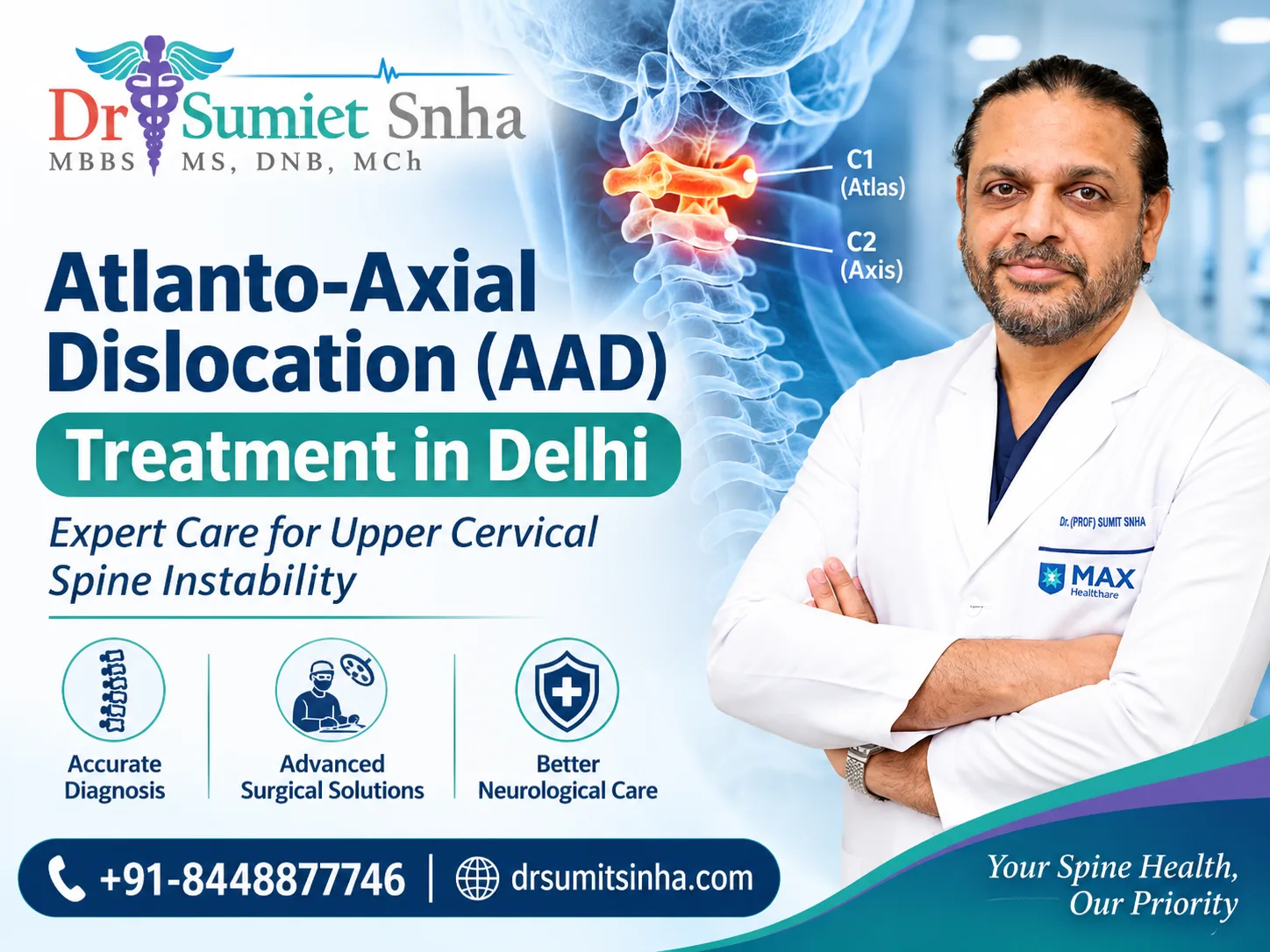
Atlanto-Axial Dislocation (AAD) Treatment in Delhi is an important consideration for patients experiencing upper neck instability, spinal cord compression, or abnormalities involving the first two cervical vertebrae. The condition requires careful evaluation because the junction between the skull and the neck contains delicate neurological structures responsible for movement, sensation and several essential bodily functions.
The upper cervical spine plays a unique role in supporting the head while allowing it to rotate and move. Any disturbance in the alignment between the first cervical vertebra (C1) and the second cervical vertebra (C2) can affect the stability of this region. Depending on the severity, patients may experience neck discomfort, restricted movement, headaches, weakness or neurological difficulties.
Understanding the condition, recognising its early symptoms and selecting an appropriate treatment approach are essential steps towards protecting spinal cord function. With modern imaging, specialised surgical techniques and individualised treatment planning, patients can receive appropriate care based on their specific spinal condition.
What Is Atlanto-Axial Dislocation?
The human cervical spine consists of seven vertebrae that support the head and protect the spinal cord. The first two vertebrae have a special anatomical structure because they work together to provide stability and rotational movement.
Atlas (C1): The uppermost cervical vertebra that supports the skull.
Axis (C2): The second cervical vertebra, which contains a tooth-like projection called the odontoid process or dens.
Transverse Ligament: A strong ligament that helps maintain the correct relationship between the atlas and axis.
Together, these structures form the atlanto-axial joint, which allows a significant portion of the rotational movement of the neck.
When the normal relationship between C1 and C2 becomes disrupted, the condition is known as Atlanto-Axial Dislocation (AAD). The displacement may occur because of congenital abnormalities, ligament damage, trauma, inflammatory disorders or other structural problems.
In certain patients, displacement can reduce the available space for the spinal cord. This may result in spinal cord compression, also known as cervical myelopathy, which can affect movement, balance and sensation.
Why Does Atlanto-Axial Dislocation Develop?
There is no single cause of atlanto-axial instability. The condition can develop during childhood, following an accident or gradually because of an underlying medical disorder.
Understanding the underlying cause is important when planning Atlanto-Axial Dislocation (AAD) Treatment in Delhi, as different forms of instability may require different treatment strategies.
1. Congenital Abnormalities
Some individuals are born with structural differences involving the craniovertebral junction. These abnormalities may remain unnoticed for years before symptoms develop.
Common congenital conditions associated with AAD include:
Os Odontoideum: A separate bone-like structure near the odontoid process that can contribute to instability.
Basilar Invagination: An upward displacement of the odontoid process towards the base of the skull.
Atlanto-Occipital Assimilation: An abnormal partial or complete fusion of the atlas with the skull.
Klippel-Feil Syndrome: A congenital condition involving fusion of two or more cervical vertebrae.
These abnormalities may alter the normal biomechanics of the upper cervical spine.
2. Traumatic Neck Injuries
Road accidents, sports injuries, falls and other significant trauma can damage the bones or ligaments supporting the C1-C2 joint.
An odontoid fracture or transverse ligament injury may cause abnormal movement between the upper cervical vertebrae. Early medical assessment is important because an unstable injury can potentially damage the spinal cord.
3. Rheumatoid Arthritis and Inflammatory Conditions
Chronic inflammatory diseases, particularly rheumatoid arthritis, can gradually weaken the ligaments and damage the joints surrounding the odontoid process.
As the supporting structures become weaker, the atlas may shift relative to the axis, increasing the possibility of spinal cord compression.
4. Infections and Other Medical Conditions
Certain infections involving the upper cervical spine can damage surrounding tissues and contribute to instability. In children, inflammation following an upper respiratory or throat infection may sometimes lead to a condition called Grisel syndrome, which involves rotatory displacement of C1 on C2.
Some skeletal disorders and connective tissue conditions can also increase the likelihood of upper cervical instability.
Symptoms That May Indicate Atlanto-Axial Instability
Symptoms depend on the amount of displacement, the duration of instability and whether the spinal cord or brainstem is affected.
Some patients initially experience mild discomfort, while others develop noticeable neurological symptoms.
Common warning signs include:
Persistent pain in the upper neck.
Headaches around the back of the head.
Difficulty rotating or moving the neck comfortably.
Abnormal head posture or a tilted neck.
Tingling or numbness in the arms, hands or legs.
Weakness in the upper or lower limbs.
Difficulty maintaining balance while walking.
Reduced hand coordination and grip strength.
Muscle stiffness or increased reflexes.
Difficulty swallowing or changes in voice in certain advanced cases.
In more serious situations, spinal cord or brainstem compression can affect essential neurological functions.
Important: New or worsening limb weakness, difficulty walking, breathing problems or bladder and bowel disturbances require urgent medical assessment. Patients with suspected upper cervical instability should avoid forceful neck manipulation, chiropractic adjustments and sudden neck movements until medically evaluated.
Different Types of Atlanto-Axial Dislocation
AAD is classified according to the direction of displacement, its stability and whether the vertebrae can return to their normal alignment.
Reducible Atlanto-Axial Dislocation
In reducible AAD, the displaced vertebrae can return towards their normal position under carefully controlled conditions. The appropriate treatment depends on the degree of instability and neurological involvement.
Irreducible Atlanto-Axial Dislocation
In this type, the displaced vertebrae cannot be brought back into their normal alignment through conventional positioning or traction. Long-standing deformity, abnormal joint anatomy or fibrous tissue may contribute to the condition.
Specialised surgical techniques may be considered to restore alignment and stabilise the affected region.
Rotatory Atlanto-Axial Dislocation
This occurs when the atlas rotates abnormally in relation to the axis. It is particularly recognised in children and may develop after minor trauma or an upper respiratory infection.
Atlanto-Axial Dislocation with Basilar Invagination
Some patients have an associated upward displacement of the odontoid process towards the skull base. This can affect the brainstem and upper spinal cord, making detailed imaging and treatment planning especially important.
How Is Atlanto-Axial Dislocation Diagnosed?
Accurate diagnosis is essential before deciding on the most suitable treatment. A specialist evaluates the patient's symptoms, medical history, neurological condition and imaging findings.
The diagnostic process may include the following investigations:
1. Clinical and Neurological Examination
The specialist evaluates neck discomfort, posture, limb strength, reflexes, coordination, walking ability and other neurological signs.
Neck movement is assessed cautiously, particularly when instability is suspected.
2. Cervical Spine X-rays
X-rays help identify abnormal alignment between C1 and C2. In selected patients, supervised dynamic imaging may be used to assess movement and instability.
Flexion-extension views should only be performed when considered safe by the treating specialist.
3. CT Scan with 3D Reconstruction
A CT scan provides detailed information about the bones of the craniovertebral junction.
It helps identify fractures, congenital abnormalities, joint deformities and the precise anatomical structure required for surgical planning.
4. MRI of the Cervical Spine
MRI helps assess the spinal cord, surrounding soft tissues, ligaments and possible compression.
It can also reveal associated abnormalities such as Chiari malformation, syringomyelia and changes within the spinal cord.
5. CT Angiography
The vertebral arteries travel close to the C1-C2 region. Their position can vary between individuals, particularly in patients with congenital abnormalities.
CT angiography may therefore be recommended before surgery to understand the vascular anatomy and plan safer screw placement.
Treatment Options for Atlanto-Axial Dislocation
Treatment depends on the underlying cause, the degree of displacement, spinal cord involvement, patient age and overall medical condition.
The objective is to maintain spinal stability, protect neurological structures and address the underlying anatomical problem.
Patients seeking Atlanto-Axial Dislocation (AAD) Treatment in Delhi should undergo an individual assessment rather than relying on a single treatment approach for every case.
1. Non-Surgical Treatment
Selected patients may be managed without surgery, particularly when the condition is stable and there is no significant spinal cord compression.
Depending on the diagnosis, conservative treatment may involve:
Appropriate medication for pain and inflammation.
Cervical collar support when indicated.
Carefully supervised immobilisation.
Treatment of associated inflammatory or infectious conditions.
Regular clinical and radiological monitoring.
Non-surgical treatment is not suitable for every patient. Significant instability, progressive neurological symptoms or spinal cord compression may require surgical intervention.
2. Surgical Stabilisation
Surgery may be recommended when the C1-C2 joint is significantly unstable, when neurological structures are compressed or when conservative management is unlikely to provide adequate stability.
The main objectives of Atlanto-Axial Dislocation (AAD) Treatment in Delhi through surgery are to restore appropriate alignment, stabilise the affected vertebrae and prevent further neurological damage.
Depending on the anatomy and type of dislocation, surgical procedures may include:
C1-C2 Posterior Fixation
This is an established surgical approach in which specialised screws and connecting rods are used to stabilise the first and second cervical vertebrae.
It helps maintain the corrected alignment and provides mechanical stability while bone fusion develops.
C1-C2 Joint Distraction and Realignment
In selected patients with irreducible dislocation or associated basilar invagination, the surgeon may use specialised techniques to open and realign the affected joints.
Bone grafts or suitable implants may be used to maintain the correction and support fusion.
Occipitocervical Fusion
When the anatomy does not permit conventional C1-C2 fixation, stabilisation may need to extend from the skull base to the upper cervical spine.
The exact fixation levels depend on the patient's anatomy and the extent of the structural abnormality.
Odontoid Screw Fixation
In selected acute odontoid fractures, an anterior screw may be used to stabilise the fracture while preserving some movement at the C1-C2 joint.
This technique is appropriate only for specific fracture patterns.
Decompression Procedures
In certain complex cases, additional decompression may be necessary when neural structures remain compressed despite realignment. The need for such a procedure depends on the individual imaging findings.
What Happens During AAD Surgery?
Surgical management of the craniovertebral junction requires detailed preparation because the spinal cord, vertebral arteries and important neural structures are located close to the operative area.
A typical surgical plan may involve the following stages:
Preoperative Assessment: Review of clinical findings, CT, MRI and other investigations.
Surgical Planning: Evaluation of bone anatomy, joint alignment and vertebral artery pathways.
Anaesthesia and Positioning: Careful positioning to minimise unnecessary movement of the unstable neck.
Surgical Exposure: Access to the required upper cervical structures through the planned approach.
Realignment: Correction of displacement when technically appropriate.
Fixation: Placement of specialised screws and rods to stabilise the affected vertebrae.
Bone Grafting: Grafting may be performed to support permanent fusion.
Postoperative Monitoring: Assessment of neurological function, wound healing and spinal alignment.
The exact procedure varies according to whether the dislocation is reducible, irreducible, traumatic or associated with congenital abnormalities.
Recovery After Atlanto-Axial Fixation Surgery
Recovery is influenced by the severity of the original condition, the duration of spinal cord compression, the surgical technique and the patient's general health.
The initial recovery period generally involves hospital monitoring, pain management and gradual mobilisation.
Early Postoperative Care
Patients are monitored for neurological changes, wound healing and other postoperative concerns. The treating team decides when sitting, standing and walking can safely begin.
Physiotherapy and Rehabilitation
A supervised rehabilitation programme may help patients improve walking ability, balance, coordination and limb strength.
Exercises should be introduced according to the surgeon's instructions, avoiding unnecessary stress on the operated region.
Follow-Up and Bone Fusion
Regular follow-up appointments and imaging help assess spinal alignment, implant position and progress towards bone fusion.
Some patients may need to wear a cervical collar for a period recommended by their surgeon.
Long-Term Activity
After C1-C2 fusion, neck rotation may be reduced because this joint normally contributes substantially to head-turning movement. Many patients gradually adapt by using other neck segments and turning their shoulders.
Return to work, driving, exercise and other daily activities should be based on clinical recovery and medical clearance.
Possible Risks and Complications
Like other complex spinal procedures, AAD surgery involves certain risks. These vary according to the patient's anatomy, medical condition and the procedure performed.
Potential complications may include:
Infection or delayed wound healing.
Bleeding or blood vessel injury.
Injury to nearby nerves or the spinal cord.
Problems with implant placement or fixation.
Failure or delay of bone fusion.
Persistent neck discomfort.
Difficulty swallowing in selected cases.
Reduced neck rotation following C1-C2 fusion.
Detailed preoperative imaging, careful surgical planning, appropriate fixation techniques and intraoperative monitoring may help reduce certain risks. However, no surgical procedure can be guaranteed to be completely risk-free.
Why Choose Dr. (Prof.) Sumiet Snha for AAD Management?
Conditions involving the craniovertebral junction require a detailed understanding of complex spinal anatomy and the relationship between the upper cervical vertebrae, spinal cord and vertebral arteries.
Dr. (Prof.) Sumiet Snha is an AIIMS-trained neurosurgeon and spine surgeon with more than 25 years of experience in neurosurgery. He has served as a Professor of Neurosurgery at AIIMS, New Delhi, and has experience in managing complex brain, spine and peripheral nerve conditions.
His professional qualifications include MBBS, MS, DNB, MCh (AIIMS, New Delhi) and FACS.
His clinical areas of expertise include:
Management of Atlanto-Axial Dislocations.
Complex craniovertebral junction conditions.
Spinal fractures and cervical spine disorders.
Minimally invasive and endoscopic spine surgery.
Spinal tumours and other complex spinal conditions.
Paediatric and adult neurosurgical care.
Dr. Snha's approach to spinal conditions involves understanding the patient's symptoms, evaluating detailed imaging and selecting a treatment strategy according to individual clinical requirements.
For more information about his professional background, qualifications and areas of expertise, visit the About Dr. (Prof.) Sumiet Snha page.
Patients looking for Atlanto-Axial Dislocation (AAD) Treatment in Delhi can seek an expert evaluation at Max Super Speciality Hospital, Dwarka, New Delhi.
Frequently Asked Questions (FAQs)
1. What is the main cause of atlanto-axial dislocation?
AAD may develop due to congenital abnormalities, traumatic injuries, ligament damage, rheumatoid arthritis, infections or other conditions affecting the upper cervical spine.
2. Is atlanto-axial dislocation a serious condition?
It can be serious, particularly when the displacement causes spinal cord or brainstem compression. Early diagnosis helps determine the risk and appropriate management.
3. Can atlanto-axial dislocation be treated without surgery?
Some stable or selected cases can be managed using immobilisation, medication and regular monitoring. Significant instability or neurological compression may require surgery.
4. What is C1-C2 fixation surgery?
C1-C2 fixation is a spinal stabilisation procedure in which screws and rods are used to hold the atlas and axis in an appropriate position. Bone grafting may also be performed to achieve permanent fusion.
5. Does AAD surgery affect neck movement?
C1-C2 fusion reduces rotational movement because the atlanto-axial joint contributes significantly to head rotation. Other cervical segments can compensate to some extent.
6. How long does recovery take after atlanto-axial surgery?
Initial recovery and hospital discharge depend on the patient's condition and surgical complexity. Bone fusion generally takes several months, while neurological recovery may continue for longer.
7. Which specialist treats atlanto-axial dislocation?
A neurosurgeon or spine surgeon experienced in craniovertebral junction disorders can evaluate the condition and recommend suitable treatment.
8. Where can I consult for atlanto-axial dislocation treatment in Delhi?
Patients can consult Dr. (Prof.) Sumiet Snha at Max Super Speciality Hospital, Dwarka, New Delhi, for an evaluation of atlanto-axial dislocation and other complex spine conditions.
Conclusion
The stability of the upper cervical spine is essential for protecting the spinal cord and maintaining normal head and neck movement. Atlanto-axial dislocation can develop for several reasons, and its effects may range from mild discomfort to significant neurological impairment.
Recognising symptoms, obtaining appropriate imaging and understanding the available treatment options can make an important difference in managing this condition. While selected patients may benefit from non-surgical care, others may require specialised stabilisation procedures to restore spinal alignment and protect neurological function.
For patients considering Atlanto-Axial Dislocation (AAD) Treatment in Delhi, consultation with an experienced neurosurgeon is an important step towards an informed treatment decision. Dr. (Prof.) Sumiet Snha provides specialised evaluation and management of complex spine and craniovertebral junction conditions at Max Hospital, Dwarka, New Delhi.

0 Comments